Observation of the hepatoprotective and antioxidant activities of Trianthema decandra Linn. (Vallai sharunnai) roots on carbon tetrachloride-treated rats
Abstract
The present study was carried out to observe the hepatoprotective effect and antioxidant activity of the ethanol extract of the roots of Trianthema decandra Linn. (200 and 400 mg/kg) in rats treated with carbon tetrachloride for 8 weeks. Extract at the tested doses restored the levels of all serum (aspartate aminotransferase, alanine aminotransferase, alkaline phosphatase, total bilirubin and total protein) and liver homogenate enzymes (glutathione peroxidase, glutathione reductase, superoxide dismutase and catalase) significantly. Histology demonstrated profound steatosis degeneration and nodule formation were observed in the hepatic architecture of carbon tetrachloride treated rats which were found to acquire near-normalcy in extract plus carbon tetrachloride administrated rats, and supported the biochemical observations. This study suggests that ethanol extract of T. decandra has a liver protective effect against carbon tetrachloride-induced hepatotoxicity and possess antioxidant activities.
Introduction
Liver, the largest organ in vertebrate body, is the major site of intense metabolic activities. Liver injury caused by toxic chemicals and certain drugs has been recognized as a toxicological problem. Herbal drugs are playing an important role in health care programs worldwide, and there is a resurgence of interest in herbal medicines for the treatment of various ailments including hepatopathy. Hepatoprotective effect of some plants like Spirulina maxima (Torres-Durain et al., 1999), Eclipta alba (Saxena et al., 1993), Boehmeria nivea (Lin et al., 1998), Cichorium intybus (Zafar and Ali, 1998) and Picrorhiza kurrooa (Saraswat et al., 1999), Phyllanthus niruri (Iqbal et al., 2007) etc has been reported. Trianthema decandra Linn. of family Aizoaceae, is a prostrate weed distributed in the Southern parts of India. The roots of the plant is well-known as an aperient (Nadkarni, 1976) and reported to be useful in hepatitis, asthma and in orchitis (Kirtikar and Basu, 1991). No scientific data have been reported for the hepatoprotective effect of this medicinal plant so far, this study was therefore undertaken to fill the lacuna in this regard.
A number of pharmacological and chemical agents act as hepatotoxin and produce variety of liver ailments (Ram and Goel, 1999). Carbon tetrachloride-intoxication in rats is an experimental model widely used to study necrotic and steatonic changes in hepatic tissue. Accordingly, the present experiment was designed to use carbon tetrachloride-intoxicated rat liver as model.
Materials and Methods
Plant material
Roots of T. decandra were collected from outskirts of Chennai, Tamil Nadu, South India. The plant was identified and authenticated by experts from the Botanical Survey of India, Southern circle, Coimbatore, Tamil Nadu, India. A voucher specimen was deposited to the Botanical Survey of India (No: BSI/SC/5/23/07-08-Tech-592).
Preparation of extract
The collected roots were washed with running water to remove sand and adherent impurities, chopped and air dried under shade. The dried roots were pulverized in an electric grinder. The powder obtained was extracted with ethanol (95% v/v). The extract was then reduced to a dark colored molten mass by using rotary evaporator under reduced pressure. The percentage yield of the extract was 14.2% with reference to the dried material. The extract suspended in 1% gum acacia was used for animal administration.
Experimental animals
Wistar rats weighing 150-200 g of either sex procured from TANUVAS (Tamil Nadu University of Veterinary and Animal Sciences), Chennai were used for the study. The animals were maintained in well ventilated rooms with 12:12 light/dark cycle in polypropylene cages. Standard rodent pellets (Hindustan Lever Ltd, Mumbai) and water were provided ad libitum. Animals were acclimatized to the laboratory conditions one week prior to the initiation of the study.
Preliminary phytochemical screening
Preliminary phytochemical screening was performed as per procedure of Kokate et al. (2004).
Acute toxicity studies
Albino mice weighing 22-25 g selected by random sampling technique were used in the study. Acute oral toxicity was performed as per OECD-423 guidelines (Ecobichon, 1997). The animals were fasted overnight, provided only water after which extract was administered to the groups orally at the dose level of 5 mg/kg body weight by gastric intubation and the groups were observed for 14 days. If mortality was observed in 2 or 3 animals, then the dose administered was assigned as a toxic dose. If mortality was observed in one animal, then the same dose was repeated again to confirm the toxic dose. If mortality was not observed, the procedure was repeated for further higher doses such as 50, 300 and 2,000 mg/kg body weight. The animals were observed for toxic symptoms such as behavioral changes, locomotion, convulsions and mortality for 72 hours.
Induction of hepatic damage
Liver damage was induced in rats by administering carbon tetrachloride (1 mL/kg) in a suspension of olive oil (1:1) orally twice a week, on every first and fourth day of all the 8 weeks (Vogel, 2002).
Experimental design
Rats were divided into 5 groups of 6 animals each as follows: Group I: Served as control and received 1 mL of 1% gum acacia p.o for 8 weeks; Group II: Served as hepatotoxic rats and received carbon tetrachloride (1 mL/kg) with equal volume of olive oil (1:1) twice a week for 8 weeks; Group III: Animals received extract (200 mg/kg p.o) suspended in 1% gum acacia for 8 weeks. Carbon tetrachloride (1 mL/kg) suspended in olive oil (1:1) was administered orally twice a week for 8 weeks concomitantly; Group IV: Animals received extract (400 mg/kg p.o) suspended in 1% gum acacia for 8 weeks. Carbon tetrachloride (1 mL/kg) suspended in olive oil (1:1) was administered orally twice a week for 8 weeks concomitantly; Group V: Animals received silymarin (25 mg/kg p.o) suspended in gum acacia (1%) for 8 weeks. Carbon tetrachloride (1 mL/kg) suspended in olive oil (1:1) was administered orally twice a week for 8 weeks.
Replenishing a known quantity of fresh food daily at 10.30 a.m. and thereby measuring the food intake of the previous day carried out measurement of daily food consumption. Body weight of rats was recorded weekly to assess percentage of weight gain in each group. General well being and behavior of the animals were observed daily throughout the period of study. The litter in the cage was renewed twice a week to ensure maximum comfort for the animals.
Animals were kept starved overnight on the 59th day. On the next day all the animals were sacrificed under light ether anesthesia. Blood was collected by carotid bleeding into sterilized dry centrifuge tubes and allowed to coagulate for 30 min at 37°C. The clear serum was separated at 2,500 rpm for 10 min, this was kept in frozen containers and preceded for biochemical estimations. A portion of liver tissue was fixed in 10% formasal (formalin diluted to 10% with normal saline) for histology. Hundred milligram of liver issue from each rat was preserved at -20°C for antioxidant study.
Biochemical estimations
The separated serum was subjected to biochemical estimation of different parameters like aspartate aminotransferase, alanine amino-transferase (Reitman and Frankel, 1957), alkaline phosphatase (Tiez, 1983); total billirubin (Gupta et al., 2007), total protein (Lowry et al., 1951) and lactate dehydrogenase (Alam, 2002).
Histology
The formasal-fixed portion of liver was processed for histopathology. After paraffin embedding and block making, serial sections of 5 µm thickness were made, stained with hemotoxylin and eosin, examined under microscope and photographed with Nikon LabPhot 2 unit (Venukumar and Latha, 2002).
The antioxidant status
One hundred milligram of liver tissue was weighed and homogenate was prepared in 10 mL Tris hydrochloric acid buffer (0.5 M; pH 7.4) at 4°C. The homogenate was centrifuged and the supernatant was used for the assay of cytoprotective enzymes namely catalase (Aebi, 1983); glutathione peroxidase (Lawrence and Burk, 1976); superoxide dismutase (Marklund and Marklund, 1974) and glutathione reductase (Dobler and Anderson, 1981).
Statistical analysis
One-way analysis of variance (ANOVA) followed by Dunnett's t-test was applied for determining the statistical significance of difference between experimental groups. The level of significance was set at 0.05.
Results
The extract did not produce any toxic symptoms of mortality up to the dose level of 2,000 mg/kg body weight in rats, and hence the drugs were considered to be safe for further pharmacological screening. According to the OECD-423 guidelines for acute oral toxicity, the LD50 dose of 2,000 mg/kg and above is categorized as unclassified.
There was a significant decrease (p<0.001) in the levels of aspartate aminotransferase, alanine aminotransferase, alkaline phosphatase and total protein and a significant increase (p<0.001) in total bilirubin in carbon tetrachloride-treated animals from those of the control group. Administration of extract (200 and 400 mg/kg) increased the reduced levels of aspartate aminotransferase, alanine aminotransferase, alkaline phosphatase and total protein (p<0.001). The level of total bilirubin was reduced significantly by the administration of extract in a dose dependent manner. Silymarin, the reference drug restored the altered levels of enzymes significantly (p<0.001) (Table I). The levels of cytoprotective enzymes such as glutathione peroxidase, the enhanced activities of these serum marker enzymes observed in tetrachloride-treated rats of the present correspond to the extensive liver damage induced by the hepatotoxin. The tendency of these enzymes to return to near normalcy in extract administered groups is a clear manifestation of antihepatotoxic effect of extract.
Table I: Effect of T. decandra on different marker enzymes and total protein
| Parameters | Group I | Group II | Group III | Group IV | Group V |
|---|---|---|---|---|---|
| Aspartate aminotransferase (IU/L) | 45.6 ± 0.6 | 121.5 ± 5.5a* | 92.3 ± 0.8b* | 65.0 ± 1.2b* | 51.7 ± 1.5b* |
| Alanine aminotransferase (IU/L) | 35.0 ± 1.0 | 101.6 ± 10.5 a* | 64.7 ± 5.1b* | 44.0 ± 1.0b* | 38.3 ± 0.7b* |
| Alkaline phosphatase (IU/L) | 76.3 ± 8.5 | 214.9 ± 11.8 a* | 155.5 ± 4.9b* | 131.0 ± 3.7b* | 118.3 ± 5.5b* |
| Total bilirubin (mg/dL) | 0.3 ± 0.03 | 2.8 ± 0.6a* | 1.3 ± 0.03 b* | 0.5 ± 0.1b* | 0.3 ± 0.03b* |
| Total protein (mg/dL) | 8.9 ± 0.2 | 3.2 ± 0.03a* | 5.4 ± 0.03 b* | 5.5 ± 0.1b* | 6.7 ± 0.2b* |
| Values are Mean ± SEM, n= 6, *p<0.001, a= Group I vs. Group II; b= Group II vs. Groups III, IV and V | |||||
Hypoalbuminemia is most frequent in the presence of advanced chronic liver diseases. Hence decline in total protein content can be deemed as a useful index of the severity of cellular dysfunction in chronic liver diseases. The lowered level of total proteins recorded in the serum as well as liver of carbon tetrachloride-treated rats suggests the severity of hepatopathy.
Table II: Effect of T. decandra on activities of antioxidant enzymes
| Parameters | Group I | Group II | Group III | Group IV | Group V |
|---|---|---|---|---|---|
| Superoxide dismutase (U/mg) | 7.8 ± 0.1 | 4.4 ± 0.1a* | 6.9 ± 0.04b* | 7.3 ± 0.1b* | 7.6 ± 0.03b* |
| Catalase (U/mg) | 88.1 ± 2.1 | 39.0 ± 2.1a* | 78.3 ± 1.5b* | 82.6 ± 1.3b* | 84.8 ± 1.3b* |
| Glutathione peroxidase (U/mg) | 8.8 ± 0.04 | 6.3 ± 0.1a* | 6.6 ± 0.04b** | 7.0 ± 0.1b** | 7.3 ± 0.2b* |
| Glutathione reductase (U/mg) | 8.5 ± 0.1 | 4.1 ± 0.04a* | 7.9 ± 0.04b* | 8.1 ± 0.1b* | 8.3 ± 0.1b* |
| Values are Mean ± SEM; n=6; *p<0.001, **p<0.01; a- Group I vs. Group II; b-Group II vs. Groups III, IV and V | |||||
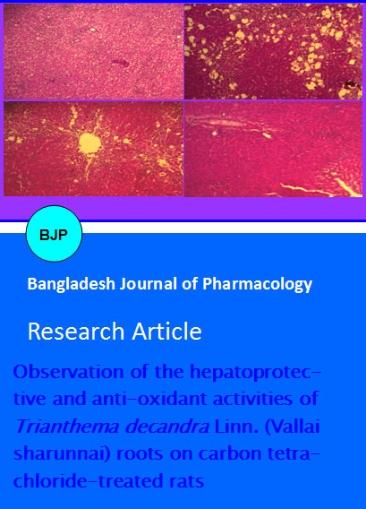
peroxidase, glutathione reductase, superoxide dismutase and catalase were reduced drastically in the liver homogenate by the administration of carbon tetrachloride plus extract (200 and 400 mg/kg) increased the levels of these enzymes (p<0.01 and p<0.001) (Table II). The histopathological sections of liver of control group and silymarin (25 mg/kg)-treated group exhibited normal architecture. The carbon tetrachloride-treated group exhibited steatosis and necrosis which was restored by the administration of extract in two different doses (Figure 1).
Figure 1: Photomicrographs of liver sections of rat stained with hemotoxylin and eosin (x100). A) normal rats (Group I) showing normal architecture; B) carbon tetrachloride-treated rats (Group II) showing steatosis and necrosis; C) carbon tetrachloride plus extract (200 mg/kg)-treated rats (Group III) showing moderate restoration of liver architecture; D) carbon tetrachloride plus extract (400 mg/kg)-treated rats (Group IV) showing almost normal of liver architecture; E) silymarin-treated rats (Group V) showing significant restoration of liver architecture comparable with normal rat
Discussion
The changes associated with carbon tetrachloride-induced liver damage of the present study appeared similar to that of acute viral hepatitis (Venukumar and Latha, 2002). Carbon tetrachloride, a widely used experimental hepatotoxicant, is biotransformed by the cytochrome P-450 system to produce the trichloromethyl free radical, which in turn covalently binds to cell membranes and organelles to elicit lipid peroxidation, disturb Ca2+ homeostasis, and finally result in cell death (Recknagel et al., 1989).
Animals of Group II (received carbon tetrachloride alone) significantly lost their body weight and showed reduced food consumption as compared to control. Animals of Group III and IV (carbon tetrachloride plus two doses of extracts) showed a significant increase in body weight and food consumption when compared to Group II. These findings suggested that extract administration has significantly neutralized the toxic effects of carbon tetrachloride and helped in regeneration of hepatocytes (Farooq et al., 1997).
Estimating the activities of serum marker enzymes, like aspartate aminotransferase, alanine aminotransferase, alkaline phosphatase can make assessment of liver function. When liver cell plasma membrane is damaged, a variety of enzymes normally located in the cytosol are released into the blood stream. Their estimation in the serum is a useful quantitative marker of the extent and glutathione reductase, superoxide dismutase and catalase. When the balance between ROS production and antioxidant defence is lost, oxidative stress results, which through a series of events deregulates the cellular functions leading to various pathological conditions (Bandyopadhyay et al., 1999). Any compound, natural or synthetic, with antioxidant properties might contribute towards the partial or total alleviation of this type of damage.
Glutathione peroxidase, superoxide dismutase and catalase constitute a mutually supportive team of defence against ROS. Superoxide dismutase is a metalloprotein and is the first enzyme involved in the antioxidant defence by lowering the steady-state level of O2-. Catalase is a hemeprotein, localized in the peroxisomes or the microperoxisomes. This enzyme catalyses the decomposition of H2O2 to water and oxygen and thus protecting the cell from oxidative damage by H2O2 and OH-. Glutathione peroxidase is a seleno enzyme two third of which (in liver) is present in the cytosol and one third in the mitochondria. It catalyses the reaction of hydroperoxidases with reduced glutathione to form glutathione disulfide (GSSG) and the reduction product of the hydroperoxide. In our study, decline in the activities of these enzymes in carbon tetrachloride-administered rats revealed that oxidative stress elicited by carbon tetrachloride intoxication have been nullified due to the effect of T. decandra. This observation agrees with those of Lin et al. (1998), who investigated hepatoprotective and antioxidant activity of Boehmeria nivea. Glutathione reductase is concerned with the maintenance of cellular level of GSH (especially in the reduced form) by effecting fast reduction of oxidized glutathione to reduced state.
It may be possible that the natural antioxidants strengthen the endogenous antioxidant defense from ROS ravage and restore the optimal balance by neutralizing the reactive species. They are gaining immense importance by virtue of their critical role in disease prevention.
Conclusion
It can be said that ethanol extract of T. decandra has exhibited a liver protective effect against carbon tetrachloride-induced hepatotoxicity and possessed antioxidant activities. Efforts are in progress to isolate and purify the active principle involved in the hepatoprotective efficacy of this medicinal plant.
Ethical Issue
The study was approved by Institutional Animal Ethical Committee (IAEC) constituted for the purpose of CPCSEA. All animal experiments were carried out according to the law of Animal Experiments Guidelines approved by National Institute of Health (NIH).
References
Aebi H. Catalase. In: Methods in enzymatic analysis. Bergmeyer HU (ed). Vol 3. New York, Academic Press, 1983, pp 276-86.
Alam H. Varley Practical clinical biochemistry. 6th ed. Gove luck, 2002, pp 522-23.
Bandyopadhyay U, Das D, Banerjee KR. Reactive oxygen species: Oxidative damage and pathogenesis. Curr Sci. 1999; 77: 658-66.
Dobler RE, Anderson BM. Simultaneous inactivation of the catalytic activities of yeast glutathione reductase by N-alkyl melimides. Biochem Biophys Acta. 1981; 70: 659.
Ecobichon DJ. The basis of toxicology testing. 2nd ed. New York, CRC Press, 1997, pp 43-60.
Farooq S, Ahmad I, Pathak GK. In vivo protective role of Koflet (an Ayurvedic preparation) against cellular toxicity caused by CCl4 and flyash. J Ethnopharmacol. 1997; 53: 109-16.
Gupta M, Mazumder UK, Thamil selvan V, Manikandan L, Senthil kumar GP, Suresh K, Kakotti BK. Potential hepatoprotective effect and antioxidant role of methanol extract of Oldenlandia umbellate in carbon tetrachloride-induced hepatotoxicity in Wistar rats. Iranian J Pharmacol Ther. 2007; 6: 5-9.
Iqbal MJ, Dewan ZF, Choudhury SAR, Mamun MIR, Mashiuzzaman M, Begum M. Pre-treatment by hexane extract of Phyllanthus niruri can alleviate paracetamol-induced damage of the rat liver. Bangladesh J Pharmacol. 2007; 2: 43-48.
Kokate CK, Purohit AP, Gokhale SB. Analytical pharmacognosy. In: Textbook of pharmacognosy. 29th ed. Pune, Nirali Prakasan, 2004, pp 108-09.
Kirtikar KR, Basu BD. Indian medicinal plants. Vol 2. 1st ed. Dehradun, International Book Distributors, 1991, p 1182.
Lawrence RA, Burk RF. Glutathione peroxidase activity in selenium deficient rat liver. Biochem Biophys Res Comm. 1976; 71: 952-58.
Lin CC, Yen MH, Lo TS, Lin JM. Evaluation of the hepatoprotective and antioxidant activity of Boehmeria nivea var. nivea and B. nivea var. tenacissima. J Ethnopharmacol. 1998; 60: 9-17.
Lowry OH, Rosebrough NJ, Farr AL, Randall RJ. Protein measurement with Folin Phenol reagent. J Biol Chem. 1951; 193: 265-75.
Marklund S, Marklund G. Involvement of superoxide anion radical in autooxidation of pyrogallol and a convenient assay of superoxide dismutase. Eur J Biochem. 1974; 47: 469-74.
Mitra SK, Venkataranganna MV, Sundaram R, Gopumadhavan S. Protective effect of HD-03, a herbal formulation, against various hepatotoxic agents in rats. J Ethnopharmacol. 1998; 63: 181-86.
Nadkarni AK. The Indian materia medica. Vol 1. 3rd ed. Bombay, Popular Prakasan, 1976, p 1228.
Prasad VS, Venkatachalam SR, Chander R, Thomas P. Dietary antioxidants-natural defense against disease. Aryavaidyan 1999; 12: 149-58.
Ram VJ, Goel A. Past and present scenario of hepatoprotectants. Curr Med Chem. 1999; 6: 217-54.
Recknagel RO, Glende EA, Jr Dolak JA, Walter RL. Mechanism of carbon tetrachloride toxicity. Pharmacol Ther. 1989; 43: 139-54.
Recknagel RO. A new direction in the study of carbon tetrachloride hepatotoxicity. Life Sci. 1983; 33: 401-08.
Reitman S, Frankel AS. A colorimetric method for the determination of serum glutamic oxaloacetic and glutamate pyruvate transaminase. Am J Clin Path. 1957; 28: 53-56.
Saraswat B, Visen PKS, Patnaik GK, Dhawan BN. Ex vivo and in vivo investigations of picroliv from Picrorhiza kurrooa and in alcohol intoxication model in rats. J Ethnopharmacol. 1999; 66: 263-69.
Saxena AK, Singh B, Anand KK. Hepatoprotective effects of Eclipta alba on sub cellular levels in rats. J Ethnopharmacol. 1993; 40: 155-16.
Tewari S, Gupta V, Bhattacharya S. Comparative study of antioxidant potential of tea with and without additives. Indian J Physiol Pharmacol. 2000; 44: 215-19.
Tiez NW. Study group on alkaline phosphatase: A reference method of measurement of alkaline phosphatase activity in human serum. Clin Chem. 1983; 29: 751.
Torres-Durain PV, Miranda-Zamora R, Paredes-Carbajal MC, Mascher D, Bie-Castillo J, Diaz-Zagoya JC. Studies on the preventive effect of Spirulina maxima on fatty liver development by carbon tetrachloride in the rat. J Ethnopharmacol. 1999; 64: 141-47.
Venukumar MR, Latha MS. Hepatoprotective effect of the methanolic extract of Curculigo orchioides in CCl4-treated male rats. Indian J Pharmacol. 2002; 34: 269-75.
Vogel GH (ed). Drug discovery and evaluation: Pharmacological assays. 2nd ed. Heidelberg, Springer-Verlag, 2002, pp 942-43.
Zafar R, Ali SM. Anti-hepatotoxic effects of root and root callus of Cichorium intybus. J Ethnopharmacol. 1998: 63: 227-31.