Tangeretin prevents prostate cancer cell proliferation and induces apoptosis via activation of Notch signalling and regulating the androgen receptor (AR) pathway and the phosphoinositide 3-kinase (PI3k)/Akt/mTOR pathways
Abstract
Despite advances in conventional therapies, treatment of prostate cancer is still a challenge. The study aims to assess the possible anti-cancer effects of tangeretin, a dietary flavonoid on human prostate cancer cells (DU145, PC3, LNCaP). Tangeretin (25, 50 and 100 µM) significantly inhibited cancer cell viability and induced DNA fragmentation and apoptosis by increasing caspase-3 and proapoptotic proteins (Bad and Bax) with reduced antiapoptotic proteins (Bcl-2 and Bcl-xL) expressions. Significant inhibition of androgen receptor (AR) and prostate specific antigen (PSA) were seen. Dose-dependent inhibition in Notch signal was observed in expression of Notch 1 and Jagged 1 along with PI3/Akt/mTOR signalling pathway. The results suggest that tangeretin inhibited prostate cancer cell proliferation and induced apoptosis via inhibiting critical pathways in cancer development - AR signalling and PI3/Akt/mTOR - Notch signalling pathways.
Introduction
Prostate cancer is one of the most common malignant cancers in men with high mortality rates (Siegel et al., 2012). Its etiology is yet to be completely understood (Bosland, 2000; Mahmoud et al., 2014) with risk factors as age, genetics, lifestyle, and diet contributing to its incidence (Whittemore et al., 1995; Crawford, 2003; Gronberg, 2003; Gupta et al., 2010). Treatment for locally confined prostate cancer is possible by surgery and/or radiation. Metastasis remains a major cause of mortality with bone metastasis in 50% of patients (Seo et al., 2006; Jemal et al., 2011) thus presenting a huge challenge to conventional therapies.
Androgen receptor signalling plays a crucial role in prostate cancer tumorigenesis (Huggins and Hodges, 1972; Chen et al., 2004; Siegel et al., 2013). Antiandrogen approaches such as androgen deprivation therapy often coupled with androgen receptor antagonists are considered as first-line therapy (Loddick et al., 2013; Schrecengost and Knudsen, 2013). Most prostate cancer respond to androgen deprivation therapy, however, patients frequently develop recurrent castration-resistant prostate cancer with aberrant reactivation of androgen receptor (Chen et al., 2004; Yuan et al., 2014). Prostate cancer progression and as well to castration-resistant prostate cancer accumulates alterations in various signalling pathways, vital to cell cycle progression and angiogenesis (Hanahan and Weinberg, 2011; Schrecengost and Knudsen, 2013).
Overexpression of pathways as the PI3K/Akt, MAPK and NFðœ…B signalling are seen in prostate cancer (Oh et al., 2001; Majumder and Sellers, 2005; Ahn et al., 2007; Polakis, 2007; Barault et al., 2008; Fecher et al., 2008). PI3K/Akt pathway is frequently activated due to loss of PTEN (phosphatase and tensin homolog) seen in around 70% prostate cancer cases (Abate-Shen and Shen, 2000; Shen and Abate-Shen, 2010).
Notch signalling pathway, vital for normal growth and development of the prostrate (Leong and Gao, 2008) regulates PTEN (Song et al., 2012). Notch is overexpressed in prostate cancer (Shou et al., 2001; Leong and Gao, 2008) and also overlaps PI3K/Akt pathway proteins (Gutierrez and Look, 2007; Salmena et al., 2008). Down-regulated Notch1 and its ligand, Jagged-1 has been shown to inhibit prostate cancer cell proliferation (Wang et al., 2010). Thus targeting Notch and PI3K/Akt pathway could possibly inhibit prostate cancer tumorigenesis.
Numerous plant-derived products have been reported to have chemopreventive properties (Yang et al., 2009; Meiyanto et al., 2012). Tangeretin, a polymethoxylated citrus flavonoid possesses biological effects as neuroprotective (Datla et al., 2001) and antimetastatic effects (Seo et al., 2011; Lakshmi and Subramanian, 2014). This study attempts to investigate the ability of tangeretin to inhibit cell proliferation and promote apoptosis in human prostate cancer cells.
Materials and Methods
Cell lines
The androgen-dependent (LNCaP) and androgen-independent (PC3 and DU145) human prostate cancer cell lines were purchased from ATCC (Chicago, USA). The cells were cultured in RPMI-1640 medium supplemented with 10% (v:v) fetal bovine serum (FBS) (Sigma Aldrich, USA), 100 μg/mL of streptomycin and 100 IU/mL of penicillin (Invitrogen Inc., USA) and incubated at 37°C with 5% CO2.
Chemicals and antibodies
Tangeretin was purchased from Sigma Aldrich (USA) and was dissolved in DMSO to obtain a stock solution of 100 mM. The desired concentrations of tangeretin were obtained by dilutions accordingly from stock solution. Primary anti-human antibodies against Bcl-2 (sc-509), Bax (sc-493), Bad (sc-8044), Bcl-xL (sc-8392), caspase-3 (sc-7148), androgen receptor (sc-7305), prostate-specific antigen (PSA) (sc-7638), PTEN (sc-7974), m-TOR (sc-8319), p-mTOR (Ser2448; sc-101738), GSK-3β (sc-9166), p-GSK-3β (sc-135653), Notch 1 (sc-9170), Jagged 1 (sc-8303) from Santa Cruz Biotechnology, USA and Akt (4685), p-Akt (Ser473, 4058) and β-actin (4970) from Cell Signaling Technology, USA were used for western blot analysis. All other chemicals used were from Sigma Aldrich (USA) otherwise are mentioned.
Determination of cell viability
The viability of prostate cancer cells were assessed as described previously (Pan et al., 2010; Zheng et al., 2010). The human prostate cancer cells were seeded in 96-well microplates at a density of 2 x 10⁵ cells/well. Cancer cells were treated with tangeretin at 25, 50 or 100 µM for 48 hours after the cells reached 70% confluence. Following incubation, 10 µL of MIT (3-(4,5-dimethylthiazol-2-yl)-2,5-diphenyltetrazolium bromide) (5 mg/mL) in PBS was added at a final concentration of 0.5 mg/mL to each well and further incubated for 4 hours. One hundred µL of a solution containing HCl (0.01 M), 10% SDS (pH 4.8) and 5% isobutyl alcohol was added to each well after discarding the supernatant and mixed well to dissolve formazan crystals. Viability of prostate cancer cells was measured at 570 nm using ELISA reader (RT6000, China). The cell viability was expressed as:
cell viability (%) = ODtest/ODcontrol x 100%
DNA fragmentation assay
DNA fragmentation following exposure to various concentrations of tangeretin was assayed by agarose gel electrophoresis as described by Wang et al. (2009). Prostate cancer cells at a density of 1 × 10⁶ cells/mL were seeded in 6-well plates and were incubated with tangeretin (25, 50 or 100 µM) for 48 hours. The cells were collected by centrifugation and total DNA was extracted using DNA isolation kit (Waston Biotechnologies Inc., China). The DNA samples were separated by electrophoresis in agarose gel (1%) and stained with ethidium bromide. The bands were detected under ultraviolet illumination by Image Master VDS-CL (Tokyo, Japan).
Western blotting
The human prostate cancer cells (PC3, DU145, and LNCaP) were incubated with tangeretin (25, 50 or 100 µM) for 48 hours. The cells were washed twice with cold PBS and lysed with lysis buffer (Cell Signalling Technology) for 20 min on ice. Cellular proteins were extracted and the concentrations were determined using BCA kit (Sigma Aldrich) according to the manufacturer’s instructions. Equal amounts of isolated protein samples were separated by electrophoresis in SDS-PAGE gels and transferred to PVDF membranes (GE Healthcare). The membranes were blocked for 1 hour at room temperature in tris-buffered saline containing 0.1% Tween-20 and 5% non-fat dry milk and were probed overnight at 4°C with primary antibodies. The membranes were then washed thrice in PBS and incubated with HRP-conjugated secondary antibodies for 1 hour at room temperature. The immunoreactive bands were visualized using enhanced chemiluminescence as described by the supplier (GE Healthcare). Band intensity was measured using ImageJ software (National Institute of Health, USA).
RNA isolation and RT-PCR
Influence of tangeretin on the expression of androgen receptor at the gene level was determined by RT-PCR analysis. Briefly, following exposure to tangeretin for 48 hours, the total cellular RNA was extracted from prostate cancer cells using TRIzol reagent (Life Technologies). cDNA was synthesized from mRNA using Thermo ScriptTM RT-PCR system (Invitrogen Inc.). The primers used were for human androgen receptor forward- GACCAGATGGCTGTCATTCA; reverse - GGAGCCATCCAAACTCTTGA, PSA forward - CATCAGGAACAAAAGCGTGA-3; reverse - AGCTG-TGGCTGACCTGAAAT and GAPDH forward - TGCAC-CACCAACTGCTTAGC; reverse - GGCATGGACTGTGG-TCATGAG. PCR reactions were carried out in eppendorf thermocycler using platinum PCR super mix (Invitrogen Inc.). PCR products were analyzed by agarose gel electrophoresis (1%) stained with ethidium bromide.
Statistical analysis
The values are expressed as mean ± SD from three or six individual experiments. The experimental data were subjected to one-way analysis of variance (ANOVA) followed by post-hoc analysis by Duncan’s Multiple Range Test (DMRT) using SPSS statistical software (version 21.0). The values at p<0.05 are considered to be statistically significant.
Results
Tangeretin inhibits prostate cancer cell proliferation
The antiproliferative effect of various concentrations of tangeretin on PC3, DU143 and LNCaP cells was assessed by MTT reduction assay. Tangeretin at 25, 50 and 100 µM concentrations caused significant (p<0.05) decline in viability of the prostate cancer cells. Concentration of 100 µM tangeretin effectively decreased viable cell counts compared to the lower doses (Figure 1). However, tangeretin induced inhibition of cell proliferation was observed to be more prominent in PC3 cells at all the concentrations as compared to LNCaP and DU145 cells. The inhibitory effects were in the order PC3>DU145>LNCaP. At 100 µM concentration, the viability percentage was 30.2%, 36.5% and 41.2% in PC3, DU145 and LNCaP cells respectively.
Figure 1: Cytotoxicity of tangeretin on human prostate cancer cells at different concentrations. Tangeretin markedly decreased the viability of the prostate cancer cells in a dose-dependent way Values are represented as mean ± SD; n=6; ‘a’ represents statistical significance at p<0.05 compared against respective controls and ‘b-e’ represents significant difference (p<0.05) between mean values within the groups of same cell line as determined by one-way ANOVA followed by DMRT analysis
Tangeretin induces DNA fragmentation of prostate cancer cells
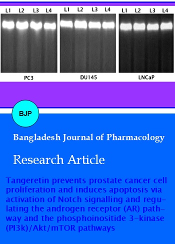
Apoptosis is a highly regulated death process that leads to the activation of endonucleases and cleavage of DNA into fragments that eventually leads to inducible non-necrotic cellular suicide. Apoptosis is pivotal in cancer therapy (Kerr et al., 1994; Piao et al., 2001) Treatment of prostate cancer cells with tangeretin resulted in fragmentation of DNA (Figure 2). Apoptosis-related fragmenting was very much visible after treatment with tangeretin at 25, 50 and 100 µM. However, 100 µM tangeretin resulted in more DNA laddering than 50 and 25 µM. DNA fragmentation was absent in cells not treated with tangeretin.
Figure 2: Tangeretin induced DNA fragmentation in prostate cancer cells. Tangeretin at different concentrations caused significant fragmentation of DNA in PC3, DU145 and LNCaP prostate cancer cells; (L1– control; L2- 25 µM tangeretin, L3- 50 µM tangeretin and L4- 100 µM tangeretin)
Tangeretin induces the expressions of apoptotic proteins
Influence of tangeretin on the expression of proteins of the apoptotic pathway was assessed by western blotting. Caspases play a crucial role as regulators of apoptosis (Zhuo et al., 2009). Tangeretin exposure bought a multi-fold increase in the expression of caspase-3. Further, as the proteins of Bcl-2 family are important in the induction of apoptosis, we also examined the effects of tangeretin on the expressions of Bcl-2 proteins. Tangeretin dose-dependently caused up-regulation in the expression of Bax and Bad with a significant (p<0.05) down-regulation of antiapoptotic proteins- Bcl-xL and Bcl-2. Tangeretin at 100 µM caused nearly two-fold enhanced expression of Bax in comparison with cells which are not exposed (Figure 3).
Figure 3: Tangeretin induced apoptosis of PCa cells by modulating the expression of the proteins of apoptotic pathway. Tangeretin induced expression of proapoptotic proteins (Bad, Bax and caspase 3) and down-regulated the expression of anti-apoptotic proteins Bcl-2 and Bcl-xL in PC 3 (a, d) DU145 (b, e) and LNCaP (c, f) cells Values are represented as mean ± SD; n=3; ‘a’ represents statistical significance at p<0.05 compared against respective controls and ‘b-e’ represents significant difference (p<0.05) between mean values within the groups of same cell line as determined by one-way ANOVA followed by DMRT analysis
Influence of tangeretin on androgen receptor and androgen receptor signalling
Studies have demonstrated that androgen-androgen receptor signalling have critical roles in prostate cancer development and progression through the transcriptional regulation of androgen receptor responsive genes in part (Lonergan and Tindall, 2011). Tumour cells also express androgens. Further, castration-resistant prostate cancers do not respond much to androgen deprivation therapy. In our study, we evaluated ability of tangeretin to inhibit the expression of androgen receptor and PSA, an important androgen receptor responsive protein and a most commonly used biomarker to indicate prostate tumor growth (Armbruster, 1993). Tangeretin potentially inhibited the expression of both androgen receptor and PSA in PC 3, DU 145 and LNCaP cells (Figure 4). Striking down-regulation of the androgen receptor and PSA was observed in cells exposed to higher concentration of tangeretin (100 µM) compared to lower doses. Furthermore, tangeretin treatment was found to have an impact at the gene expression levels, that was evident by the drastic (p<0.05) reduction in expressions of androgen receptor and PSA mRNA in all the prostate cancer cells (Figure 4). These observations suggest that tangeretin was effective in down-regulating androgen receptor as well as androgen receptor-signalling path-way.
Figure 4: Tangeretin regulates androgen receptor and androgen receptor-signalling. Tangeretin inhibits expressions of androgen receptor and PSA at mRNA levels (a) as determined by RT-PCR and also western blotting analysis reveals that tangeretin down-regulates the expressions of androgen receptor (b, d) and PSA (c, e) at protein level Values are represented as mean ± SD, n=3. “a†represents statistical significance at p<0.05 compared against respective controls and “b-e†represents significant difference (p<0.05) between mean values within the groups of same cell line as determined by one-way ANOVA followed by DMRT analysis. (L1– control; L2- 25 µM tangeretin; L3- 50 µM tangeretin and L4- 100 µM tangeretin)
Effect of tangeretin on PI3/Akt signalling cascade
The PI3K/AKT pathway that is crucial in cell survival is activated in various cancers including prostate cancer (Wang et al., 2015). Accumulating reports indicate possible crosstalk between PI3K/Akt/mTOR and androgen receptor signalling that directly regulates androgen receptor expression and activation (Morgan et al., 2009). Inhibition of the pathway could also contribute to down-regulation of androgen receptor expression. Tangeretin (25, 50 and 100 µM) doses effectively decreased expression of Akt, p-Akt, p-GSK3β and mTOR with up-regulation of PTEN expression levels (Figure 5). Loss of PTEN function is reported in nearly 30-50% of CaP (Morgan et al., 2009). The strikingly elevated expressions of PTEN observed following tangeretin exposure suggests the efficacy of tangeretin in down-regulating the PI3/Akt signalling cascades.
Figure 5: Tangeretin inhibits the PI3K/Akt/mTOR signalling. Tangeretin potentially inhibited the PI3K/Akt/mTOR signalling pathway as evidenced by the significant down-regulation of Akt, mTOR and GSK 3β with enhanced levels of PTEN
Influence of tangeretin on Notch signalling
Notch signalling plays an important role in cell fate determination in embryonic and adult tissues (Gridley, 2007; Fortini, 2009) and normal prostatic development as well as in pathogenesis of prostate cancers (Leong and Gao, 2008). Notch signalling has been reported to be abnormally activated in cancers (Carvalho et al., 2014). Marked down-regulation in the expression of Notch 1 was observed following tangeretin exposure.
Tangeretin significantly (p<0.05) inhibited the expression levels of Notch 1 ligand, Jagged 1 in the prostate cancer cells (Figure 6). Further, tangeretin at 25 µM though decreased the expression of Notch and Jagged 1 but the effects were non-significant. Concentration of 50 and 100 µM brought about significant (p<0.05) changes in all the 3 prostate cancer cell lines.
Figure 6: Tangeretin effectively modulated Notch signalling in prostate cancer cells. Tangeretin inhibits expression of Notch 1 (a, c) and (b, d) Values are represented as mean ± SD; n=3; ‘a’ represents statistical significance at p<0.05 compared against respective controls and ‘b-e’ represents significant difference (p<0.05) between mean values within the groups of same cell line as determined by one-way ANOVA followed by DMRT analysis
Discussion
Tangeretin effectively inhibited the viability of PC3, DU145 and LNCaP cells. Further, it also caused fragmentation of DNA which is an important feature of cells undergoing apoptosis (Chen et al., 1996; Yu et al., 1999). Induction of apoptosis could be a valuable strategy in successful cancer treatment.
Apoptosis, a programmed cell death occurs via activation of the cell suicide machinery (Kaufmann and Hengartner, 2001). Apoptosis is one of the main goals of anticancer therapies (Farnebo et al., 2010; Nouri and Yazdanparast, 2011). Apoptotic cascade is activated via a complex interactions of molecular events (Pulido and Parrish, 2003) of extrinsic (death receptor-dependent) and intrinsic (mitochondria-dependent) pathways (Thornberry et al., 1997).
To further assess the effects of tangeretin on the molecular events of apoptosis, the expression of Bcl-2 family proteins were determined following tangeretin exposure. The Bcl-2 family members are chief players of the mitochondria-dependent intrinsic pathway of apoptosis.The Bcl-2 family consists of proapoptotic (Bax, Bid and Bad) and antiapoptotic proteins (Bcl-2 and Bcl-xL) that maintain the balance between cell survival and death (Gross et al., 1999; Reed, 2001; Zhao et al., 2003). Bax promotes the proapoptotic factors and induce apoptosis, while antiapoptotic protein, Bcl-2 inhibits the release of proapoptotic factors and prevents apoptosis (Yang et al., 2006). The down-regulated expression of Bcl-2 and Bcl-xL along with enhanced expression of Bax and Bad observed on exposure to tangeretin possibly contributes to the tangeretin-induced apoptosis of prostate cancer cells. Activation of executioner caspases subsequently leads to apoptosis. In our study, up-regulated expression of caspase-3 was observed in prostate cancer cells upon tangeretin treatment suggesting induction of caspase cascade leading to apoptosis. The observations of the study suggest that tangeretin-induced decrease in cell viability could be due to inhibition of cell division and induction of apoptosis as well.
The prostate cells depend on androgen-mediated-androgen receptor signalling for growth, development and function which also gets retained in most of the prostate cancers (Heinlein and Chang, 2004). Constitutive androgen receptor signalling leads to the translation of chief genes involved in tumorigenesis (Wendel et al., 2007) and also in castration-resistant prostate cancer, most of the prostate cancer cells express androgen receptor, suggesting the crucial role of androgen receptor signalling in the progression of prostate cancer (Heinlein and Chang, 2004). While studies report elevated androgen receptor expression in progression to castration-resistant prostate cancer, experiments with rodent models demonstrate de novo androgen receptor up-regulation in prostate cancer (Stanbrough et al., 2001; Zhu et al., 2011). Thus targeting the androgen receptor signalling is vital for treatment, where androgen deprivation therapy is not much effective. RT-PCR analysis following exposure to various concentrations of tangeretin indicated down-regulation in the levels of androgen receptor mRNA correlated with decrease in androgen receptor expression as analysed by western blotting. Also, the expression levels of PSA, one of the androgen- responsive genes is a clinically important marker for prostate cancer (Kupelian et al., 1996; Sato et al., 1996), was also found reduced in line with androgen receptor indicating effective down-regulation and blocking of the androgen receptor-signalling pathway by tangeretin. Thus, tangeretin could possibly stand as an effective source of treatment of castration-resistant prostate cancers. Exploring further to gain insights on molecular mechanisms of inhibition of androgen receptor-signalling pathway by tangeretin would be valuable in cancer therapy.
The PI3K/Akt/mTOR cascade, plays critical roles in mammalian cell survival signalling and is the most frequently altered signalling pathway in various human cancers and it controls various processes involved in oncogenesis such as cell cycle, motility, metabolism, angiogenesis, genomic instability etc., (Morgan et al., 2009; Sarker et al., 2009; Fruman and Rommel, 2014). PI3K/Akt-signalling pathway is constitutively activated in prostate cancer (Majumder and Sellers, 2005; Carnero, 2010). Many proteins of the cascade are significantly altered in prostate cancer as PTEN, Akt and mTOR (Taylor et al., 2010; Lonigro et al., 2011). The hyperactivation of Akt and loss of function of the tumor suppressors such as PTEN have been observed in PCs (Obata et al., 1998; Trotman et al., 2006). This hyperactivation of PI3K/Akt sequentially leads to invasive carcinoma (Wang et al., 2003; Blando et al., 2009; Sircar et al., 2009). This makes PI3K/Akt signalling pathway as a potential candidate in therapy. Further it has been reported that activation of the PI3K/Akt pathway is involved in acquired resistance (Sunters et al., 2003; Zhong et al., 2010; Wang et al., 2013). Observations of our study showed significantly up-regulation of PTEN with markedly down-regulated expression of Akt, mTOR and p-GSK3β. The reduced levels of p-mTOR and p-GSK3β may possibly be due to the suppression of levels of Akt and p-Akt in prostate cancer cells exposed to tangeretin. Also it has been reported that PI3K and androgen receptor reciprocally regulate functions (Carver et al., 2011). However, in our study activation of PI3K and androgen receptor signalling pathways were observed that were markedly down-regulated by tangeretin. Preclinical assessments of dual inhibition of PI3K and androgen receptor reported antiproliferative effects (Carver et al., 2011). Thus the inhibition of androgen receptor and PI3K/Akt pathway suggests the potency of tangeretin as anti-cancer compound.
Furthermore, many studies have reported that Notch 1 signalling regulates PTEN expression in prostate tumors (Palomero et al., 2007; Whelan et al., 2009; Wang et al., 2010).
Notch signalling interacts with the androgen receptor and PI3k/Akt pathways, the mechanisms that are important in prostate growth, homeostasis and carcinogenesis (Sarker et al., 2009; Shen and Abate-Shen, 2010). Several studies have demonstrated high-level constitutive expression of Notch 1 in human prostate cancer cell lines (PC3, DU145, and LNCaP) (Shou et al., 2001; Bin et al., 2009). Gene silencing of Notch 1 suppresses malignant properties such as invasion (Bin et al., 2009), survival and proliferation (Zhang et al., 2006). Santagata et al. (2004) reported that Notch ligand, Jagged 1 expression was associated with prostate cancer metastasis and recurrence. Further down-regulation of Notch 1 and Jagged 1 resulted in inhibition of cell growth, migration, and also induced apoptosis via inactivation of Akt, mTOR, NF-κB, MMP-9 and uPA signalling pathways in prostate cancer cells (Bin et al., 2009; Wang et al., 2010). The observed down-regulation in Notch 1 and Jagged 1 following tangeretin exposure indicates the capacity of tangeretin in inhibiting prostate cancer cell growth and metastasis.
Conclusion
The results of the study reveal the potency of tangeretin in prostate cancer treatment. Inhibition of androgen receptor and androgen receptor-signalling along with inhibition of crucial pathways in prostate cancer- Notch and PI3K/Akt/mTOR suggests tangeretin as a potent candidate in therapy of prostate cancer and castration-resistant prostate cancers as well. Tangeretin is thus valuable in combined treatment strategies.
References
Abate-Shen C, Shen MM. Molecular genetics of prostate cancer. Genes Dev. 2000; 14: 2410-34.
Ahn KS, Sethi G, Aggarwal BB. Nuclear factor-𜅠B: From clone to clinic. Current Molecular Medicine 2007; 7: 619-37.
Armbruster DA. Prostate-specific antigen: Biochemistry, analytical methods, and clinical application. Clin Chem. 1993; 39: 181-95.
Barault L, Veyrie N, Jooste V, Lecorre D, Chapusot C, Ferraz JM, Lièvre A, Cortet M, Bouvier AM, Rat P, Roignot P, Faivre J, Laurent-Puig P, Piard F. Mutations in the RASMAPK, PI(3)K (phosphatidylinositol-3-OH kinase) signalling network correlate with poor survival in a population-based series of colon cancers. Int J Cancer. 2008; 122: 2255-59.
Bin HB, Adhami VM, Asim M, Siddiqui IA, Bhat KM, Zhong W, Saleem M, Din M, Setaluri V, Mukhtar H. Targeted knockdown of Notch1 inhibits invasion of human prostate cancer cells concomitant with inhibition of matrix metalloproteinase-9 and urokinase plasminogen activator. Clin Cancer Res. 2009; 15: 452-59.
Blando J, Portis M, Benavides F, Alexander A, Mills G, Dave B, Conti CJ, Kim J, Walker CL. PTEN deficiency is fully penetrant for prostate adenocarcinoma in C57BL/6 mice via mTOR-dependent growth. Am J Pathol. 2009; 174: 1869-79.
Bosland MC. The role of steroid hormones in prostate carcinogenesis. J Natl Cancer Inst Monogr. 2000; 27: 39-66.
Carnero A. The PKB/AKT pathway in cancer. Curr Pharm Des. 2010; 16: 34-44.
Carvalho FL, Simons BW, Eberhart CG, Berman DM. Notch signaling in prostate cancer: A moving target. Prostate 2014; 74: 933-45.
Carver BS, Chapinski C, Wongvipat J, Hieronymus H, Chen Y, Chandarlapaty S, Arora VK, Le C, Koutcher J, Scher H, Scardino PT, Rosen N, Sawyers CL. Reciprocal feedback regulation of PI3K and androgen receptor signalling in PTEN-deficient prostate cancer. Cancer Cell. 2011; 19: 575-86.
Chen X, Ko LJ, Jayaraman L, Prives C. p53 levels, functional domains, and DNA damage determine the extent of the apoptotic response of tumor cells. Genes Dev. 1996; 10: 2438-51.
Chen CD, Welsbie DS, Tran C, Baek SH, Chen R, Vessella R, Rosenfeld MG, Sawyers CL. Molecular determinants of resistance to antiandrogen therapy. Nat Med. 2004; 10: 33-39.
Crawford E. Epidemiology of prostate cancer. Urology 2003; 62: 3-12.
Datla KP, Christidou M, Widmer WW, Rooprai HK, Dexter DT. Tissue distribution and neuroprotective effects of citrus flavonoid tangeretin in a rat model of Parkinson’s disease. Neuroreport 2001; 12: 3871-75.
Farnebo M, Bykov VJ, Wiman KG. The p53 tumor suppressor: A master regulator of diverse cellular processes and therapeutic target in cancer. Biochem Biophys Res Commun. 2010; 396: 85-89.
Fecher LA, Amaravadi RK, Flaherty KT. The MAPK pathway in melanoma. Curr Opinion Oncol. 2008; 20: 183-89.
Fortini ME. Notch signalling: The core pathway and its posttranslational regulation. Dev Cell. 2009; 16: 633-47.
Fruman DA, Rommel C. PI3K and cancer: Lessons, challenges and opportunities. Nat Rev Drug Discov. 2014; 13: 140-56.
Gridley T. Notch signaling in vascular development and physiology. Development 2007; 134: 2709-18.
Gronberg H. Prostate cancer epidemiology. Lancet 2003; 361: 859-64.
Gross A, McDonnell JM, Korsmeyer SJ. Bcl-2 family members and the mitochondria in apoptosis. Genes Dev. 1999; 13: 1899-911.
Gupta S, Kim J, Prasad S, Aggarwal B. Regulation of survival, proliferation, invasion, angiogenesis, and metastasis of tumor cells through modulation of inflammatory pathways by nutraceuticals. Cancer Metastasis Rev. 2010; 29: 405-34.
Gutierrez A, Look AT. NOTCH and PI3K-AKT pathways intertwined. Cancer Cell. 2007; 12: 411-13.
Hanahan D, Weinberg RA. Hallmarks of cancer: The next generation. Cell 2011; 144: 646-74.
Heinlein CA, Chang C. Androgen receptor in prostate cancer. Endocr Rev. 2004; 25: 276-308.
Huggins C, Hodges CV. Studies on prostatic cancer I the effect of castration, of estrogen and androgen injection on serum phosphatises in metastatic carcinoma of the prostate. CA Cancer J Clin. 1972; 22: 232-40.
Jemal A, Bray F, Center MM, Ferlay J, Ward E, Forman D. Global cancer statistics. CA: Cancer J Clin. 2011; 61: 69-90.
Kaufmann SH, Hengartner MO. Programmed cell death: Alive and well in the new millennium. Trends Cell Biol. 2001; 11: 526-34.
Kerr JF, Winterford CM, Harmon BV. Apoptosis: Its significance in cancer, and cancer therapy. Cancer 1994; 73: 2013-26.
Kupelian P, Katcher J, Levin H, Zippe C, Klein E. Correlation of clinical and pathologic factors with rising prostate-specific antigen profiles after radical prostatectomy alone for clinically localized prostate cancer. Urology 1996; 48: 249-60.
Lakshmi A, Subramanian S. Tangeretin, a citrus pentamethoxyflavone, exerts cytostatic effect via p53/p21 up-regulation and suppresses metastasis in 7, 12-dimethylbenz [a] anthracene induced rat mammary carcinoma. J Nutri Biochem. 2014; 25: 1140-53.
Leong KG, Gao WQ. The Notch pathway in prostate development and cancer. Differentiation 2008; 76: 699-716.
Loddick SA, Ross SJ, Thomason AG, Robinson DM, Walker GE, Dunkley TP, Brave SR, Broadbent N, Stratton NC, Trueman D, Mouchet E, Shaheen FS, Jacobs VN, Cumberbatch M, Wilson J, Jones RD, Bradbury RH, Rabow A, Gaughan L, Womack C, Barry ST, Robson CN, Critchlow SE, Wedge SR, Brooks AN. AZD3514: A small molecule that modulates androgen receptor signaling and function in vitro and in vivo. Mol Cancer Ther. 2013; 12: 1715-27.
Lonergan PE, Tindall DJ. Androgen receptor signaling in prostate cancer development and progression. J Carcinog. 2011; 10: 20.
Lonigro RJ, Grasso CS, Robinson DR, Jing X, Wu YM, Cao X, Quist MJ, Tomlins SA, Pienta KJ, Chinnaiyan AM. Detection of somatic copy number alterations in cancer using targeted exome capture sequencing. Neoplasia 2011; 13: 1019-25.
Mahmoud AM, Yang W, Bosland MC. Soy isoflavones and prostate cancer: A review of molecular mechanisms. J Steroid Biochem Mol Biol. 2014; 140: 116-32.
Majumder PK, Sellers WR. Akt-regulated pathways in prostate cancer. Oncogene 2005; 24: 7465-74.
Meiyanto E, Hermawan A, Anindyajati. Natural products for cancer-targeted therapy: Citrus flavonoids as potent chemopreventive agents. Asian Pac J Cancer Prev. 2012; 13: 427-36.
Morgan TM, Koreckij TD, Corey E. Targeted therapy for advanced prostate cancer: Inhibition of the PI3K/Akt/mTOR pathway. Curr Cancer Drug Targets. 2009; 9: 237-49.
Nouri K, Yazdanparast R. Proliferation inhibition, cell cycle arrest and apoptosis induced in HL-60 cells by a natural diterpene ester from Daphne mucronata. Daru 2011; 19: 145-53.
Obata K, Morland SJ, Watson RH, Hitchcock A, Chenevix-Trench G, Thomas EJ, Campbell IG. Frequent PTEN/MMAC mutations in endometrioid but not serous or mucinous epithelial ovarian tumors. Cancer Res. 1998; 58: 2095-97.
Oh AS, Lorant LA, Holloway JN, Miller DL, Kern FG, El-Ashry D. Hyperactivation of MAPK induces loss of ER𛼠expression in breast cancer cells. Mol Endocrinol. 2001; 15: 1344-59.
Palomero T, Sulis ML, Cortina M, Real PJ, Barnes K, Ciofani M, Caparros E, Buteau J, Brown K, Perkins SL, Bhagat G, Agarwal AM, Basso G, Castillo M, Nagase S, Cordon-Cardo C, Parsons R, Zúñiga-Pflücker JC, Dominguez M, Ferrando AA. Mutational loss of PTEN induces resistance to NOTCH1 inhibition in T-cell leukemia. Nat Med. 2007; 13: 1203-10.
Pan MH, Lin CL, Tsai JH, Ho CT, Chen WJ. 3,5,3’,4’,5’-Pentamethoxystilbene (MR-5), a synthetically methoxylated analogue of resveratrol, inhibits growth and induces G1 cell cycle arrest of human breast carcinoma MCF-7 cells. J Agric Food Chem. 2010; 58: 226-34.
Piao W, Yoo J, Lee DK, Hwang HJ, Kim JH. Induction of G2/M phase arrest and apoptosis by a new synthetic anticancer agent, DW2282, in promyelocytic leukemia (HL-60) cells. Biochem Pharmacol. 2001; 62: 1439-47.
Polakis P. The many ways of Wnt in cancer. Curr Opinion Genetics Develop. 2007; 17: 45-51. Pulido MD, Parrish AR. Metal-induced apoptosis: Mechanisms Mutat Res. 2003; 533: 227-41.
Reed JC. Apoptosis-regulating proteins as targets for drug discovery. Trends Mol Med. 2001; 7: 314-19.
Salmena L, Carracedo A, Pandolfi PP. Tenets of PTEN tumor suppression. Cell 2008; 133: 403-13.
Santagata S, Demichelis F, Riva A, Varambally S, Hofer MD, Kutok JL, Kim R, Tang J, Montie JE, Chinnaiyan AM, Rubin MA, Aster JC. JAGGED1 expression is associated with prostate cancer metastasis and recurrence. Cancer Res. 2004; 64: 6854-57.
Sarker D, Reid AH, Yap TA, de Bono JS. Targeting the PI3K/AKT pathway for the treatment of prostate cancer. Clin Cancer Res. 2009; 15: 4799-805.
Sato N, Gleave ME, Bruchovsky N, Rennie PS, Goldenberg SL, Lange PH, Sullivan LD. Intermittent androgen suppression delays progression to androgen-independent regulation of prostate-specific antigen gene in the LNCaP prostate tumour model. J Steroid Biochem Mol Biol. 1996; 58: 139-46.
Schrecengost RS, Knudsen KE. Molecular pathogenesis and progression of prostate cancer. Seminars in Oncology 2013; 40: 244-58.
Seo Y, Franc BL, Hawkins RA, Wong KH, Hasegawa BH. Progress in SPECT/CT imaging of prostate cancer. Technol Cancer Res Treatment 2006; 5: 329-36.
Seo J, Lee HS, Ryoo S, Seo JH, Min BS, Lee JH. Tangeretin, a citrus flavonoid, inhibits PGDF-BB-induced proliferation and migration of aortic smooth muscle cells by blocking AKT activation. Eur J Pharmacol. 2011; 673: 56-64.
Shen MM, Abate-Shen C. Molecular genetics of prostate cancer: new prospects for old challenges. Genes Dev. 2010; 24: 1967-2000.
Shou J, Ross S, Koeppen H, de Sauvage FJ, Gao WQ. Dynamics of Notch expression during murine prostate development and tumorigenesis. Cancer Res. 2001; 61: 7291-97.
Siegel R, Desantis C, Virgo K, Stein K, Mariotto A, Smith T, Cooper D, Gansler T, Lerro C, Fedewa S, Lin C, Leach C, Cannady RS, Cho H, Scoppa S, Hachey M, Kirch R, Jemal A, Ward E. Cancer treatment and survivorship statistics, 2012. CA Cancer J Clin. 2012; 62: 220-41.
Siegel R, Naishadham D, Jemal A. Cancer statistics, 2013. CA Cancer J Clin. 2013; 63: 11-30.
Sircar K, Yoshimoto M, Monzon FA, Koumakpayi IH, Katz RL, Khanna A, Alvarez K, Chen G, Darnel AD, Aprikian AG, Saad F, Bismar TA, Squire JA. PTEN genomic deletion is associated with p-Akt and AR signalling in poorer outcome, hormone refractory prostate cancer. J Pathol. 2009; 218: 505-13.
Song MS, Salmena L, Pandolfi PP. The functions and regulation of the PTEN tumour suppressor. Nat Rev Mol Cell Biol. 2012; 13: 283-96.
Stanbrough M, Leav I, Kwan PW, Bubley GJ, Balk SP. Prostatic intraepithelial neoplasia in mice expressing an androgen receptor transgene in prostate epithelium. Proc Natl Acad Sci USA. 2001; 98: 10823-28.
Sunters A, Fernández de Mattos S, Stahl M, Brosens JJ, Zoumpoulidou G, Saunders CA, Coffer PJ, Medema RH, Coombes RC, Lam EW. FoxO3a transcriptional regulation of Bim controls apoptosis in paclitaxel-treated breast cancer cell lines. J Biol Chem. 2003; 278: 49795-805.
Taylor BS, Schultz N, Hieronymus H, Gopalan A, Xiao Y, Carver BS, Arora VK, Kaushik P, Cerami E, Reva B, Antipin Y, Mitsiades N, Landers T, Dolgalev I, Major JE, Wilson M, Socci ND, Lash AE, Heguy A, Eastham JA, Scher HI, Reuter VE, Scardino PT, Sander C, Sawyers CL, Gerald WL. Integrative genomic profiling of human prostate cancer. Cancer Cell. 2010; 18: 11-22.
Thornberry NA, Rano TA, Peterson EP, Rasper DM, Timkey T, Garcia-Calvo M, Houtzager VM, Nordstrom PA, Roy S, Vaillancourt JP, Chapman KT, Nicholson DW. A combinatorial approach defines specificities of members of the caspase family and granzyme B. Functional relationships established for key mediators of apoptosis. J Biol Chem. 1997; 272: 17907-11.
Trotman LC, Alimonti A, Scaglioni PP, Koutcher JA, Cordon-Cardo C, Pandolfi PP. Identification of a tumour suppressor network opposing nuclear Akt function. Nature 2006; 441: 523-27.
Wang S, Gao J, Lei Q, Rozengurt N, Pritchard C, Jiao J, Thomas GV, Li G, Roy-Burman P, Nelson PS, Liu X, Wu H. Prostate-specific deletion of the murine Pten tumor suppressor gene leads to metastatic prostate cancer. Cancer Cell. 2003; 4: 209-21.
Wang H, Xu Y, Yan J, Zhao X, Sun X, Zhang Y, Guo J, Zhu C. Acteoside protects human neuroblastoma SH-SY5Y cells against beta-amyloid-induced cell injury. Brain Res. 2009; 4: 139-47.
Wang Z, Li Y, Banerjee S, Kong D, Ahmad A, Nogueira V, Hay N, Sarkar FH. Down-regulation of Notch-1 and Jagged-1 inhibits prostate cancer cell growth, migration and invasion, and induces apoptosis via inactivation of Akt, mTOR, and NF-kB signalling pathways. J Cell Biochem. 2010; 109: 726-36.
Wang H, Galbán S, Wu R, Bowman BM, Witte A, Vetter K, Galban CJ, Ross BD, Cho KR, Rehemtulla A. Molecular imaging reveals a role for AKT in resistance to cisplatin for ovarian endometrioid adenocarcinoma. Clin Cancer Res. 2013; 19: 158-69.
Wang P, Phan T, Gordon D, Chung S, Henning SM, Vadgama JV. Arctigenin in combination with quercetin synergistically enhances the antiproliferative effect in prostate cancer cells Mol Nutr Food Res. 2015; 59: 250-61.
Wendel HG, Silva RL, Malina A, Mills JR, Zhu H, Ueda T, Watanabe-Fukunaga R, Fukunaga R, Teruya-Feldstein J, Pelletier J, Lowe SW. Dissecting eIF4E action in tumorigenesis. Genes Dev. 2007; 21: 3232-37.
Whelan JT, Kellogg A, Shewchuk BM, Hewan-Lowe K, Bertrand FE. Notch-1 signaling is lost in prostate adenocarcinoma and promotes PTEN gene expression. J Cell Biochem. 2009; 107: 992-1001.
Whittemore AS, Kolonel LN, Wu AH, John EM, Gallagher RP, Howe GR. Prostate cancer in relation to diet, physical activity, and body size in blacks, whites, and Asians in the United States and Canada. J Natl Cancer Inst. 1995; 87: 652- Yang SH, Chien CM, Lu MC, Lin YH, Hu XW, Lin SR. Upregulation of Bax and endonuclease G, and down-modulation of Bcl-XL involved in cardiotoxin III-induced apoptosis in K562 cells. Exp Mol Med. 2006; 38: 435-44.
Yang C, Wang X, Lu G, Picinich S. Cancer prevention by tea: Animal studies, molecular mechanisms and human relevance. Nat Rev Cancer. 2009; 9: 429-39.
Yu Z, Chen J, Ford BN, Brackley ME, Glickman BW. Human DNA repair systems: An overview. Environ Mol Mutagen. 1999; 33: 3-20.
Yuan X, Cai C, Chen S, Chen S, Yu Z, Balk SP. Androgen receptor functions in castration-resistant prostate cancer and mechanisms of resistance to new agents targeting the androgen axis. Oncogene 2014; 33: 2815-25.
Zhang Y, Wang Z, Ahmed F, Banerjee S, Li Y, Sarkar FH. Down-regulation of Jagged-1 induces cell growth inhibition and S phase arrest in prostate cancer cells. Int J Cancer. 2006; 119: 2071-77.
Zhao H, Yenari MA, Cheng D, Sapolsky RM, Steinberg GK. Bcl-2 overexpression protects against neuron loss within the ischemic margin following experimental stroke and inhibits cytochrome c translocation and caspase-3 activity. J Neurochem. 2003; 85: 1026-36.
Zheng P, Liu YX, Chen L, Liu XH, Xiao ZQ, Zhao L, Li GQ, Zhou J, Ding YQ, Li JM. Stathmin, a new target of PRL-3 identified by proteomic methods, plays a key role in progression and metastasis of colorectal cancer. J Proteome Res. 2010; 9: 4897-905.
Zhong B, Sallman DA, Gilvary DL, Pernazza D, Sahakian E, Fritz D, Cheng JQ, Trougakos I, Wei S, Djeu JY. Induction of clusterin by AKT–role in cytoprotection against docetaxel in prostate tumor cells. Mol Cancer Ther. 2010; 9: 1831-41.
Zhu C, Luong R, Zhuo M, Johnson DT, McKenney JK, Cunha GR, Sun Z. Conditional expression of the androgen receptor induces oncogenic transformation of the mouse prostate. J Biol Chem. 2011; 286: 33478-88.
Zhuo L, Gong J, Yang R, Sheng Y, Zhou L, Kong X, Cao K. Inhibition of proliferation and differentiation and promotion of apoptosis by cyclin L2 in mouse embryonic carcinoma P19 cells. Biochem Biophys Res Commun. 2009; 18: 451-57.