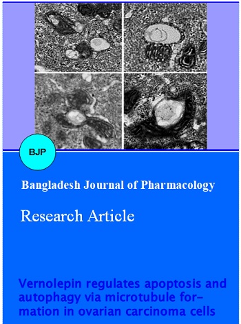
Vernolepin regulates apoptosis and autophagy via microtubule formation in ovarian carcinoma cells
Abstract
The study demonstrates the effect of vernolepin on proliferation and apoptosis in ovarian cancer cell lines. The inhibition of cell growth was significant at 30 μM concentration after 48 hours in both OVCAR-3 and SK-OV3 cell lines. Phase-contrast microscopic examination revealed a decrease in number of vernolepin-treated cells. A number of membranous structures and vacuoles were visible in the cytoplasm after 24 hours. After 48 hours chromatin condensation and nuclear fragmentation indicating typical apoptotic changes were observed. Vernolepin treatment lead to 83.6% cell viability compared to control. However the cell viability was increased to 93.7% on after starvation followed by vernolepin treatment. On the other hand, 3-MA in combination with vernolepin decreased cell viability to 54.5%. Annexin V-FITC/PI staining and FACS demonstrated that in OVCAR-3 and SK-OV3 cells treatment with vernolepin (30 µΜ) for 48 hours caused apoptosis in 34.2% and 28.5% cells respectively. Thus, vernolepin-treatment in ovarian carcinoma cells leads to autophagy before the onset of apoptosis and protects cancer cells.
Introduction
In United States out of gynaecologic cancers, ovarian cancer is the leading cause of death (Chan et al., 2006). The lifetime risk of ovarian cancer for women with family history in the US is estimated 9.4% (Hartge et al., 1994). Absence of symptoms during early stage, leads detection of ovarian cancer at metastatic stage (Van et al., 2000). Only 25% of the epithelial ovarian cancers are detected as Stage I of the disease (Bast, 2003). The currently available treatment regimens and cytoreductive surgery has increased the cure rate of cancer patients at Stage I to 90% (Hoskins, 1995). No doubt use of surgery followed by chemotherapy has increased the response rates (Eisenkop et al., 2003; McGuire et al., 1995; Ozols et al., 2003) but complications associated with disease progression cause 75% deaths afterwards. Although, cisplatin is the most important antineoplastic agent against a wide variety of solid tumors (Previati et al., 2006) but the development of drug resistance hinders its efficiency (Stewart et al., 2006). Thus the demand for novel therapeutic agents is unmet.
Autophagy plays a vital role in response to some physiological processes (Terman et al., 2007) and may lead to type II programmed cell death (Baehrecke, 2005). In case of many antitumor drugs activation of autophagy leads to inhibition of cell proliferation. It has been observed in rats with carcinogen-induced pancreatic cancer that pancreatic adenocarcinoma cells have lower autophagic activity during tumor progression (Toth et al., 2002). During starvation autophagy acts as a mechanism of cell survival. Autophagy is activated as a defence mechanism in response to many cancer therapeutics (Wu et al., 2008).
Vernolepin is a sesquiterpene lactone isolated from the dried fruits of Vernonia amygdalina (Laekeman et al., 1983). It shows platelet anti-aggregating properties (Laekeman et al., 1985) and is also an irreversible DNA polymerase inhibitor (Clayde, 2005) hence may have antitumor properties. In the present study, we investigated the effect of vernolepin on autophagy and cell death in ovarian carcinoma cells.
Materials and Methods
Reagent
Vernolepin, acridine orange (AO), monodansylcadervarine (MDC), 4,6-diamidino-2-phenylindole (DAPI), 3-methyladenine (3-MA) and rabbit polyclonal antibody against LC3 were purchased from Sigma-Aldrich (USA). Lipofectamine 2000 was obtained from Invitrogen (Carlsbad, CA, USA) and Annexin V-FITC Kit from Bender MedSystems (Vienna, Austria).
Cell lines and culture
OVCAR-3 and SK-OV3 cell lines were obtained from The Cell Bank of Type Culture Collection of Chinese Academy of Sciences, Shanghai Institute of Cell Biology (China). The cells were maintained in Dulbecco’s modified Eagle medium supplemented with 10% fetal bovine serum containing penicillin 100 units/mL, streptomycin 100 mg/mL and 10% FBS (PAA).
CCK-8 assay
In 96-well plates 1 × 10⁵ cells/well were distributed. The plates after incubation for 12 hours were treated with either DMSO or different doses of vernolepin for different time periods. To each well 10 μL of CCK-8 solution (Dojindo Laboratories) was added. The plates were incubated for 1 hour followed by measurement of absorbance at 450 nm using a microplate spectrophotometer (BIO-RAD xMark).
DAPI staining
In 96-well culture plates the cells treated with vernolepin were fixed with 4% paraformaldehyde for 20 min at 37°C. The cells were then stained with DAPI for 20 min and observed under the fluorescence microscope (OLYMPUS IX71).
Analysis of apoptosis and cell cycle arrest using FACS
The OVCAR-3 and SK-OV3 cells onto 6-well plates were treated with DMSO or vernolepin for 24 or 48 hours after incubation for 12 hours. The cells at a density of 3 x 10⁵/mL were washed in PBS and suspended in binding buffer. Annexin V-FITC (5 μL) was added to 200 μL cell suspension, incubated for 20 min, washed and resuspended in 200 μL binding buffer. Addition of 10 μL propidium iodide [PI] (20 μg/mL) was followed by FAC Scan analysis for apoptotic cell death. Cells fixed with 75% ice-cold ethanol at -20°C for 2 hours were stained with PI in the presence of RNase A (100 μg/mL) for cell cycle analysis.
AO or MDC vital staining
In 96-well plates, the cells treated with vernolepin were stained with AO (5 μg/mL) or MDC (0.05 mmol/L) for 20 min. We used fluorescence microscope (OLYMPUS IX71) for examination of the stained cells.
Western blot analysis
Following vernolepin treatment, the cells were lysed in lysis buffer (150 µmol/L NaCl, 50 µmol/L Tris-HCl, 0.5% deoxycholic acid, 1% NP-40, 0.1% SDS and protease inhibitor cocktail) on ice for 1 hour. Bicinchoninic acid protein assay kit (Pierce) was used for determination of protein concentration after centrifugation of lysates. Proteins separated on SDS-PAGE were transferred to polyvinylidenedifluoride (PVDF) membrane blocked with a solution containing 10 µmol/L Tris, 150 µmol/L NaCl, 0.1% Tween 20 (TBS-T) and 5% non-fat dry milk for 3 hours. After incubation with primary antibody at 4°C for 12 hours the membrane was treated with secondary antibody for 2 hours.
Statistical analysis
We used student’s t distribution probability density function for calculation of P-value. The experiments were performed in triplicates and expressed as mean ± S.D. The differences were considered statistically significant at p<0.05.
Results
Treatment of human ovarian carcinoma cell lines with vernolepin lead to inhibition of cell growth in a concentration and time dependent manner. Among the range of concentrations from 10 to 100 μM tested the inhibition of cell growth was significant at 30 μM of vernolepin after 48 hours in both OVCAR-3 and SK-OV3 cell lines (Figure 1A).
Analysis of cell cycle using PI staining and FACS showed an increase in the number of OVCAR-3 cells at G2/M phase (18.2 ± 1.9% to 39.2 ± 2.1%) on vernolepin treatment after 48 hours. Similar cell cycle arrest was also detected in SK-OV3 cells (15.0 ± 1.0% to 47.7 ± 2.4%) at G2/M phase (Figure 1B).
Figure 1: Vernolepin inhibited the proliferation of ovarian cancer cells; (B) Cell cycle was arrested at G2/M phase by vernolepin. The data are presented as means ± S.D. from three independent experiments
Although no change in nuclei or nuclear membrane was noticed on vernolepin treatment in OVCAR-3 cells but a number of membranous structures and vacuoles were visible in the cytoplasm after 24 hours. After 48 hours chromatin condensation and nuclear fragmentation indicating typical apoptotic changes was clearly visible (Figure 2A).
There was only cytoplasmic vacuolization with no chromatin condensation in the vernolepin-treated SK-OV3 cells for 24 hours. Autophagosomes were also observed in the cytoplasm, which contained portions of cytosol and organelles, such as endoplasmic reticulum (ER) and mitochondria (Figure 2B).
Figure 2: Formation of autophagosomes was induced by vernolepin in ovarian cancer cells. OVCAR-3 (A); and SK-OV3 (B) cells were treated with vernolepin (30 μM) or DMSO
In vernolepin-treated OVCAR-3 cells red fluorescent spots whereas in control cells and the cells co-treated with 5 µmol/L 3-MA plus vernolepin green fluorescence was observed on AO staining. In vernolepin-treated OVCAR-3 cells accumulation of autophage specific staining MDC was also observed around the nuclei (Figure 3).
Figure 3: Accumulation of AVOs induced by vernolepin treatment
OVCAR-3 cells on starvation (in EBSS medium) for 4 hours, exhibited reduction in cell viability to 66.9%. Similarly treatment of OVCAR-3 cells with 5 µmol/L 3-MA decreased cell viability to 76.2% (Figure 4). Vernolepin treatment lead to 83.6% cell viability compared to control. However the cell viability was increased to 93.7% on treatment with 2 hour starvation followed by 48 hours vernolepin treatment. On the other hand, 3-MA (2 µmol/L) in combination with vernolepin decreased cell viability to 54.5% (Figure 4). These results demonstrate that in vernolepin-treated OVCAR-3 cells autophagy exerts protective effect.
Figure 4: Protective role of autophagy in vernolepin-treated OVCAR-3 cells
The results from DAPI staining revealed typical apoptotic changes with chromatin condensation and nuclear fragmentation in OVCAR-3 cells at 48 hours of vernolepin treatment. Annexin V-FITC/PI staining and FACS demonstrated that in OVCAR-3 cells treatment with 30 μM vernolepin for 48 hours caused 34.2% of the cells to undergo apoptosis compared to 3.5% in control. Similarly treatment with 30 μM vernolepin for 48 hours caused 28.5% SK-OV3 cells to undergo apoptosis compared to 2.7% in control (Figure 5).
Figure 5: Vernolepin induced apoptosis in ovarian cancer cells after 48 hours treatment
Discussion
The present study demonstrates that vernolepin treatment inhibits cell proliferation, arrests G2/M phase of cell cycle and induces the formation of AVOs in OVCAR-3 and SK-OV3 cancer cells. We also observed a large number of double-membrane autophagosomes and PAS in both OVCAR-3 and SK-OV3 ovarian cancer cell after vernolepin treatment. In addition, ovarian cancer cells on co-treatment with 3-MA and vernolepin, showed that 3-MA could reverse the accumulation of MDC and AO. All these results indicated that 3-MA could specifically inhibit vernolepin-induced autophagy in ovarian cancer cells by inhibiting the increase of LC3.
Recently, radiation (Paglin et al., 2001), ceramide (Daido et al., 2004), rapamycin (Takeuchi et al., 2005), and arsenic trioxide (Kanzawa et al., 2003) have been reported to act as stimulants for the autophagic process. The results from our study revealed that autophagy could play a protective role in vernolepin-treated ovarian cancer cells. Nutrient starvation has been widely used to induce autophagy (Martinez-Borra and Lopez-Larrea, 2012). OVCAR-3 cells on co-treatment with starvation and vernolepin exhibited high level of autophagy compared to the cells treated with vernolepin alone. However, there was increased vernolepin-induced cell death on treatment with 3-MA. Vernolepin treatment for 48 hours significantly increased apoptosis in OVCAR-3 and SK-OV3 cells. There was chromatin condensation, nuclear fragmentation and disappearance of the surface microvilli in the cells undergoing apoptosis.
Conclusion
Vernolepin treatment could induce autophagy in ovarian cancer cells. Autophagy could protect cancer cells from apoptotic cell death by delaying the onset of apoptosis. P21 protein plays an important role in vernolepin-induced autophagy and apoptosis.
References
Baehrecke EH. Autophagy: Dual roles in life and death? Nat Rev Mol Cell Biol. 2005; 6: 505-10.
Bast RC Jr. Status of tumor markers in ovarian cancer screening. J Clin Oncol. 2003; 21: 200s-05s.
Chan JK, Cheung MK, Husain A, Teng NN, West D, Whittemore AS, Berek JS, Osann K. Patterns and progress in ovarian cancer over 14 years. Obstet Gynecol. 2006; 108: 521-28.
Clayde J. Organic chemistry. Oxford, Oxford University Press, 2005, p 238.
Daido S, Kanzawa T, Yamamoto A, Takeuchi H, Kondo Y, Kondo S. Pivotal role of the cell death factor BNIP3 in ceramide-induced autophagic cell death in malignant glioma cells. Cancer Res. 2004; 64: 4286-93.
Eisenkop SM, Spirtos NM, Friedman RL, Lin WC, Pisani AL, Perticucci S. Relative influences of tumor volume before surgery and the cytoreductive outcome survival for patients with advanced ovarian cancer: A prospective study. Gynecol Oncol. 2003; 90: 390-96.
Hartge P, Whittemore AS, Itnyre J, McGowan L, Cramer D. Rates and risks of ovarian cancer in subgroups of white women in the United States. The Collaborative ovarian Cancer Group. Obstet Gynecol. 1994; 84: 760-64.
Hoskins WJ. Prospective on ovarian cancer: Why prevent? J Cell Biochem. 1995; 23(Suppl): 189-99.
Kanzawa T, Kondo Y, Ito H, Kondo S, Germano I. Induction of autophagic cell death in malignant glioma cells by arsenic trioxide. Cancer Res. 2003; 63: 2103-08.
Laekeman GM, Mertens J, Totté J, Bult H, Vlietinck AJ, Herman AG. Isolation and pharmacological characterization of vernolepin. J Nat Prod. 1983; 46: 161–69.
Laekeman GM, Clerck F, Vlietinck AJ, Herman AG. Vernolepin: An antiplatelet compound of natural origin. Naunyn-Schmiedeberg's Arch Pharmacol. 1985; 331: 108–13.
Martinez-Borra J, Lopez-Larrea C. Autophagy and self-defense. Adv Exp Med Biol. 2012; 738: 169-84.
McGuire WP, Hoskins WJ, Brady MF, Kucera PR, Partridge EE, Look KY, Clarke-Pearson DL, Davidson M. Cyclophosphamide and cisplatin compared with paclitaxel and cisplatin in patients with stage III and stage IV ovarian cancer. N Engl J Med 1996; 334: 1-6.
Ozols RF, Bundy BN, Greer BE, Fowler JM, Clarke-Pearson D, Burger RA, Mannel RS, DeGeest K, Hartenbach EM, Baergen R. Phase III trial of carboplatin and paclitaxel compared with cisplatin and paclitaxel in patients with optimally resected stage III ovarian cancer: A Gynecologic Oncology Group study. J Clin Oncol. 2003; 21: 3194-200.
Paglin S, Hollister T, Delohery T, Hackett N, McMahill M, Sphicas E, Domingo D, Yahalom J. A novel response of cancer cells to radiation involves autophagy and formation of acidic vesicles. Cancer Res. 2001; 61: 439-44.
Previati M, Lanzoni I, Corbacella E, Magosso S, Guaran V, Martini A, Capitani S. Cisplatin-induced apoptosis in human promyelocytic leukemia cells. Int J Mol Med. 2006; 18: 511–16.
Stewart JJ, White JT, Yan X, Collins S, Drescher CW, Urban ND, Hood L, Lin B. Proteins associated with Cisplatin resistance in ovarian cancer cells identified by quantitative proteomic technology and integrated with mRNA expression levels. Mol Cell Proteomics. 2006; 5: 433–43.
Terman A, Gustafsson B, Brunk UT. Autophagy, organelles and ageing. J Pathol. 2007; 211: 134-43.
Toth S, Nagy K, Palfia Z, et al. Cellular autophagic capacity changes during azaserine-induced tumour progression in the rat pancreas: Up-regulation in all premalignant stages and down-regulation with loss of cycloheximide sensitivity of segregation along with malignant transformation. Cell Tissue Res. 2002; 309: 409-16.
Takeuchi H, Kondo Y, Fujiwara K, et al. Synergistic augmentation of rapamycin-induced autophagy in malignant glioma cells by phosphatidylinositol 3-kinase/protein kinase B inhibitors. Cancer Res. 2005; 65: 3336-46.
Van Dalen A, Favier J, Burges A, Hasholzner U, de Bruijn HW, Dobler-Girdziunaite D, Dombi VH, Fink D, Giai M, McGing P, Harlozinska A, Kainz C, Markowska J, Molina R, Sturgeon C, Bowman A, Einarsson R. Prognostic significance of CA 125 and TPS levels after 3 chemotherapy courses in ovarian cancer patients. Gynecol Oncol. 2000; 79: 444-50.
Wu YT, Tan HL, Huang Q, Kim YS, Pan N, Ong WY, Liu Z, Ong CN, Shen HM. Autophagy plays a protective role during zVAD-induced necrotic cell death. Autophagy 2008; 4: 457-66.

