Computational analyses on delineating specificity of 2-hydroxy-3,5-dinitrobenzamide, a BH3-mimetic, towards antiapoptotic proteins
Abstract
We have herein computationally examined binding affinities and specificity of 2-hydroxy-3,5-dinitrobenzamide (HDNB), a small chemical molecular BH3-mimetic identified by means of "peptidodynmimetic method", on the BH3-binding grooves of six antiapoptotic proteins (Bcl-2, Bcl-B, Bcl-W, Bcl-XL, Bfl-1 and Mcl-1) from human beings. The HDNB ligand was found to interact on the BH3-binding grooves of Bcl-2, Bcl-B, Bcl-XL, Bfl-1 and Mcl-1, whereas it did not act as BH3-mimetic to Bcl-W. Moreover, binding affinities of the HDNB towards the antiapoptotic proteins were significantly different from each other. The differential binding affinities and specificity of the HDNB towards the antiapoptotic proteins have been chiefly attributed to the differences in the chemical properties of BH3-binding grooves of the proteins. Implications of the study to design efficient de novo antagonists to the antiapoptotic proteins using the HDNB as seed molecule have been discussed.
Introduction
Apoptosis is tightly regulated by intrinsic pathway activated by Bcl-2 family proteins present in the mitochondrial membrane and antiapoptotic proteins of the Bcl-2 family are important cancer biomarkers as they are over expressed in all types of cancers (Kelly and Strasser, 2011). The antiapoptotic proteins are also playing essential roles in various biological activities: the Bcl-2 and the Bcl-XL are playing essential roles for regulating B-lymphocytes and platelet survival, respectively; similarly, Mcl-1 and Bcl-W are essential for the sustainable production of hematopoetic stem cells and sperm cells, respectively (Lessene et al., 2008). Therefore, administration of anti-cancer drugs interacting with antiapoptotic proteins exhibiting no over expression may presumably cause some adverse effect (such as lymphopenia, thrombocytopaenia and anemia) to cancer patients. In these contexts, examining binding affinities and specificities of antagonists to the antiapoptotic proteins is crucial to avoid/reduce late-stage attritions in drug discovery processes.
We have recently reported 2-hydroxy-3,5-dinitrobenzamide (HDNB) as a drug-likeness inhibitor to human Bcl-B (hBcl-B) by using an array of computational strategies (Sivakumar et al., 2013). However, binding preferences and specificity of the HDNB towards other five antiapoptotic proteins have been left unaddressed to date.
In the present study, we have studied binding affinities of the HDNB with each antiapoptotic protein using molecular docking methods and also stringently scrutinized mode of interactions of the lead with the proteins. Moreover, implications of the study to design highly potent de novo inhibitors to the antiapoptotic proteins have also been discussed.
Materials and Methods
Selections and preparations of antiapoptotic proteins: Experimental three-dimensional (3D) structures of all the six antiapoptotic proteins Bcl-2 (1GJH), Bcl-B (4B4S), Bcl-W (1ZY3), Bcl-XL (2YXJ), Bfl-1 (3MQP) and Mcl-1 (2KBW) were retrieved from protein data bank . Missing hydrogen atoms were added and bond orders were assigned to all proteins considered, herein, by using Schrodinger suite 9.2 and hydrogen bonds were also defined to the structures based on sample water orientation. Using PROPKA module of the Schrodinger suite 9.2, the protein structures were optimized to pH 7.0. The resultant structures were energy minimized using "Steepest descent" algorithm with a tolerance of 1000 KJ/mol/nm and step size of 0.01 with required number of minimization steps. The energy minimized structures were subjected to molecular dynamics (MD) simulations in near physiological conditions (pH 7.0, 1 atmospheric pressure and ionic strength of 0.1 M NaCl) for 5 ns at 310 K by Gromacs 4.5.1 (prior to productive runs, each protein was equilibrated for 200 ps and 500 ps under NVT and NPT conditions, respectively). In each case, trajectory structures were stored at every 25 ps and the data were analyzed in terms of change in potential energy and RMSD of backbone atoms of the proteins. The data analyses revealed that all proteins could attain equilibrium under the defined conditions well within the time span of 5 ns MD. A representative structure from the equilibrium phase of each antiapoptotic protein was selected and used as 3D structures of the proteins for all computational studies performed in the present studies unless otherwise mentioned.
Molecular dockings and analyses of non-covalent interactions in the interfaces of antiapoptotic proteins- HDNB complexes: 3D structure of 2-hydroxy-3,5-dinitrobenzamide (ID: MMs01085173) were downloaded from MMsINC database. Using the OPLS2005 force field, all possible ionization states of the compound were processed at pH 7.0 using Ionizer algorithm in the ligand preparation module of Schrodinger suite. The program was allowed to generate all combinations of stereoisomers and all pH-dependent chemical forms of the HDNB. The program was allowed to generate low energy ring conformation (with limitation of one per ligand) of the compound. Grid boxes (having dimensions of 80 x 80 x 80 Angstrom) were set to fully cover the BH3 binding grooves (composed of helices 2-5) of the antiapoptotic proteins. All generated structures of the HDNB were docked on the BH3-binding grooves of the 6 antiapoptotic proteins by Glide-XP with default parameters except that total numbers of poses per run, total numbers of generated poses per ligand for molecular docking and selected poses per ligand in the post-docking processes were set as 10,000, 100 and 10, respectively. Scaling factor and partial charge cut-off were set to 0.8 and 0.15, respectively, for all molecular docking runs of the present study. Various non-covalent interactions for each of the antiapoptotic – HDNB complexes were analyzed using Schrodinger suite 9.2 and PyMol 1.5.0.1 molecular visualization tools.
Result and Discussion
There are 6 antiapoptotic proteins from human being as reported in the literature to date. They are named as Bcl-2, Bcl-B, Bcl-W, Bcl-XL, Bfl-1 and Mcl-1. All the antiapoptotic proteins are single polypeptide chains composed of about 250 standard amino acids and they are simple helical proteins (Ke et al., 2001). The antiapoptotic proteins belonging to Bcl-2 family have similar 3D structures depicting well-defined BH3-binding groove (Liang and Fesik, 1997). All the antiapoptotic proteins are playing essential roles on regulating/resisting apoptosis. Moreover, each of the proteins plays unique roles in various biological processes, notwithstanding similar 3D structures of the proteins (Placzek et al., 2010). However, exact structure-activity relationships of the proteins have not yet well addressed to our best knowledge. As these antiapoptotic proteins are retarding apoptosis upon inactivating pro-apoptotic proteins and are also over expressed in cancer diseases, designing of small molecular inhibitors to the proteins has become an important strategy in cancer chemotherapy (Labi et al., 2008). Many drug-likeness chemical inhibitors to the antiapoptotic proteins have been reported by means of experimental/computational methods in the literature (Sivakumar et al., 2012). In these contexts, we have recently reported 2-hydroxy-3,5-dinitrobenzamide (HDNB) as a novel BH3-mimetic to hBcl-B using pepti-dodynmimetic method (Sivakumar et al., 2013). In the present study, specificity of the HDNB towards the antiapoptotic proteins has been demonstrated in detail.
Two-dimensional representation of HDNB structure is shown in Figure 1. The molecule has a six-member ring substituted by four charged chemical moieties. The ligand was docked on the BH3-binding grooves of the antiapoptotic proteins and the docking studies suggested that the HDNB was capable to interact with all the antiapoptotic proteins. However, the HDNB did not interact on the BH3-binding groove of Bcl-W, but did with all other five proteins. GlideScores calculated for the HDNB to interact on the BH3-binding groove of Bcl-2, Bcl-B, Bcl-XL, Bfl-1 and Mcl-1 were ranged from 3.1 to 4.1 implying that the HDNB has moderate differences in its binding preferences towards the proteins (Table I). The HDNB showed relatively stronger interactions with Bcl-XL and Mcl-1 comparing its interactions with Bcl-B, Bcl-2 and Bfl-1. The differential binding affinities of the HDNB is intriguing as the BH3-binding groove of each antiapoptotic protein is unanimously constituted by residues from helices 2, 3, 4 and 5 of the respective proteins (Bernardo et al., 2008; Bernardo et al., 2010; Bernardo et al., 2011; Schreiber and Keating, 2011). However, the BH3-binding grooves of Bcl-W and Mcl-1 are significantly wider comparing the counterpart regions in Bcl-2, Bcl-B, Bcl-XL and Bfl-1. Moreover, binding locations and orientations of the HDNB on the BH3-binding grooves of the 5 antiapoptotic proteins were also different from each other (Figure 1).
Table I: Docking parameters and residues of various non-covalent interactions for antiapoptotic proteins-HDNB complexes
| Proteins and Pdb ID | Glide score | LEb | Residues within 4 Angstrom to the ligand | Hydrogen bonds* | Salt bridgea |
|---|---|---|---|---|---|
| Bcl-2 (1GJH) | 3.2 | 0.20 | F104,Y108, D111, F112, M115,Q118, V133, L137, A149, F153 | OE1(Q118)....H3(HDNB) OD1(D111)....H5(HDNB) OD2(D111).....H5(HDNB) |
OD1(D111)....N2(HDNB) |
| Bcl-B (4B4S) | 3.3 | 0.20 | L46, H50, F54, M71, V88, T89, T92, F93 | ND1(H50)....H4(HDNB) | - |
| Bcl-W (1ZY3) | 2.9 | 0.18 | Q80, V81, D83, E84, Q87, T89 | O(Q80)....H4(HDNB) OE2(E84)....H5(HDNB) |
OE2(E84)....N2(HDNB) |
| Bcl-XL (2YXJ) | 4.1 | 0.25 | A93, E96, F97, R100, Y101,W137, G138, V141, F191, Y195 | - | - |
| Bfl-1 (3MQP) | 3.1 | 0.20 | V48, N51, L52, V74, M75, K77, E78, T91, F95 | O(K77)....H3(HDNB) OE1(E78)....H4(HDNB) |
- |
| Mcl-1 (2KBW) | 3.8 | 0.24 | H224, A227, F228, M231, V249, H252, V253, S255, T266, L267, V270 | O(H252)....H4(HDNB) | - |
| aVarious atoms of amino acids are represented by standard nomenclature; H3 (HDNB) and H4 (HDNB) denote amide protons of the HDNB; H5 (HDNB) denotes hydroxyl proton of the HDNB; N2 (HDNB) denotes nitrogen atom of a nitro group present in the HDNB; aLigand efficiency (Rajesh et al., 2013) | |||||
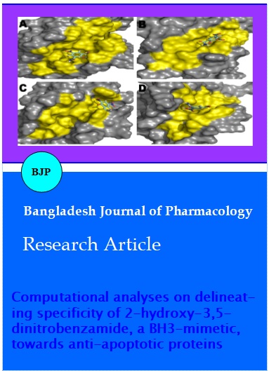
Figure 1: Docking complexes of HDNB and antiapoptotic proteins [A = Bcl-2, B = Bcl-B, C = Bcl-XL, D = Bfl-1, E = Mcl-1, G = Bcl-W (front view of BH3-binding groove) and H = Bcl-W (rear view of BH3-binding groove)]. The antiapoptotic proteins (in similar orientations) and HDNB are shown in surface and sticks models, respectively. The BH3-binding grooves of the antiapoptotic proteins are painted in yellow color for sake of clarity. F = Two dimensional structure of HDNB (2-hydroxy-3,5-dinitrobenzamide) drawn by using ChemSketch is shown
Amide protons of the HDNB were invariably in close proximity to any of the residues such as aspartic acid, glutamic acid, glutamine, histidine and lysine located in the BH3-binding grooves of the antiapoptotic proteins: amide protons in the HDNB could be observed to have hydrogen bond interaction with Gln118, His50, Lys77 and His252 of Bcl-2, Bcl-B, Bfl-1 and Mcl-1, respectively. In the case of Bcl-XL, amide protons of the HDNB were found to be about 3.1 Angstrom and 4.0 Angstrom away from backbone carbonyl group and side-chain carbonyl group of Glu96 of the protein, respectively. These interactions were not classified as hydrogen bonds (H-bonds) as the distance cutoff used in the Schrodinger suite 9.2 for the present study was 2.5 Angstrom for defining inter molecular H-bonds. Similarly, hydroxyl group of the HDNB was found to be in close contacts with the residues such as Asp111, Thr89, Tyr101, Thr91 and His224 located in the BH3-binding grooves of Bcl-2, Bcl-B, Bcl-XL, Bfl-1 and Mcl-1, respectively. While the amide and hydroxyl groups of the HDNB were showed electrostatic interactions with charged and polar amino acids, rest of the ligand portion was surrounded by bulky hydrophobic amino acids such as leucine, methionine, phenylalanine and valine present in the BH3-binding grooves of the antiapoptotic proteins (Table I). It should be mentioned that the HDNB was unable to bind on the BH3-binding groove of Bcl-W, which was constituted by residues such as Ala48, Phe52 (from helix 2) Leu63 (from helix 3), Arg77, Phe78, Val81, Ser82 (from helix 4) and Trp92, Val96, Ala97, Ala100, Phe101, Tyr150 (from helix 5). Strikingly, the BH3- binding groove of the Bcl-W was remarkably different from the counterpart regions of other 5 antiapoptotic proteins: The BH3-binding grooves of the 5 proteins are composed of about 60% hydrophobic amino acids and rest of regions in the grooves of the proteins was found to be occupied with charged and polar amino acids. In contrary, the groove of Bcl-W was predominantly (about 90%) constituted by hydrophobic amino acids. The surface groove of the Bcl-W was found to have only an arginine and a serine residue at positions of 77 and 82, respectively (there were no acidic amino acids). Thus, the differential binding affinities of the HDNB towards Bcl-W and other antiapoptotic proteins could be presumably rationalized to differences in chemical properties of the BH3-binding grooves of the respective proteins. On the other hand, the HDNB showed relatively weak interaction (vis-a -vis its interaction with other proteins) on a surface groove composed of residues such as Gln80, Val81, Asp83, Glu84, Gln87 and Thr89 of the hBcl-W (Table I and Figure 1) implying that the HDNB was not acting as BH3-mimetic to the antiapoptotic protein.
Though the six antiapoptotic proteins are homologous and have similar three-dimensional structures, over expression patterns of the proteins in cancers are not identical (Yip and Reed, 2008). Moreover, structural stabilities and biological activities of the proteins are also different from each other (Reed, 2008). As demonstrated herein and reported in the literature, binding affinities of the six antiapoptotic proteins towards lead anti-cancer compounds and as well to BH3-only peptides are different from each other (Sivakumar et al., 2011; Sivakumar and Sivaraman, 2012). And, in the present study, the HDNB depicted differential binding affinities and specificity towards 6 antiapoptotic proteins whose BH3-binding grooves are different from each other in terms of sizes, shapes and chemical properties (as discussed above). On the other hand, an amide group and a hydroxyl group positioned at 1st and 2nd positions of the HDNB (Figure 1) are crucial for interacting with all the 6 antiapoptotic proteins (though the ligand did not bind on the BH3-binding groove of the Bcl-W, Table I and Figure 1). In addition, six member ring of the ligand has two nitro groups at 3rd and 5th positions, which were found to involve for establishing salt-bridge interactions only in Bcl-2 - HDNB and Bcl-W - HDNB complexes. In other words, the two nitro groups did not make any essential non-bonding interactions with other 4 antiapoptotic proteins (Bcl-B, Bcl-XL, Bfl-1 and Mcl-1). In these backgrounds, structural determinants identified for the HDNB and as well for antiapoptotic proteins for generating protein-ligand complexes may pave ways for developing highly efficient and specific prototype molecules to each of the antiapoptotic proteins using HDNB as seed molecule by means of "core hopping" (Li et al., 2011) and "de novo" (Sivakumar and Sivaraman, 2011; Rajesh and Sivaraman, 2013) drug designing strategies in near future.
Conclusion
Differential interactions of the HDNB towards the six antiapoptotic proteins have been chiefly attributed to the differences in the chemical properties of the BH3-binding grooves of the proteins and in turn the findings may be useful on designing highly specific de novo antagonists to each of the antiapoptotic proteins.
Acknowledgements
The authors would like to acknowledge the Centre of Excellence – AYUSH at SASTRA University, for generously allowing us to use Schrodinger software suite supported by the AYUSH research grant (Z.15015/01/2010), Government of India.
References
Bernardo PH, Wan K, Sivaraman T, Xu J, Moore FK, Hung AW, Mok HYK, Yu VC, Chai CLL. Structure-Activity Relationship Studies of phenanthridine-based Bcl-XL inhibitors. J Med Chem. 2008; 51: 6699-710.
Bernardo PH, Sivaraman T, Wan K, Xu J, Krishnamoorthy J, Sons CM, Tian L, Chin JSF, Lim DSW, Mok HYK, Yu VC, Tong JC, Chai CLL. Structural insights into the design of small molecule inhibitors that selectively antagonize mcl-1. J Med Chem. 2010; 53: 2314-18.
Bernardo PH, Sivaraman T, Wan KF, Xu J, Krishnamoorthy J, Song CM, Tian L, Chin JS, Lim DS, Mok HY, Yu VC, Tong JC, Chai CL. Synthesis of a rhodanine-based compound library targeting Bcl-XL and Mcl-1. Pure Appl Chem. 2011; 83: 723-31.
Ke N, Godzik A, Reed JC. Bcl-B, a Novel Bcl-2 family member that differentially binds and regulates Bax and Bak. J Biol Chem. 2001; 276: 12481-84.
Kelly PN, Strasser A. The Role of Bcl-2 and its pro-survival relatives in tumourigenesis and cancer therapy. Cell Death Differ. 2011; 18: 1414-24.
Labi V, Grespi F, Baumgartner F, Villunger A. Targeting the Bcl-2-regulated apoptosis pathway by BH3 mimetics: A breakthrough in anticancer therapy? Cell Death Differ. 2008; 15: 977-87.
Lessene G, Czabotar PE, Colman PM. Bcl-2 family antagonists for cancer therapy. Nat Rev Drug Discov. 2008; 7: 989-1000
Li XB, Wang SQ, Xu WR, Wang RL, Chou KC. Novel inhibitor design for hemagglutinin against H1N1 influenza virus by core hopping method. PLoS One. 2011; 6: e28111.
Liang H, Fesik SW. Three-dimensional structures of proteins involved in programmed cell death. J Mol Biol. 1997; 274: 291-302.
Placzek WJ, Wei J, Kitada S, Zhai D, Reed JC, Pellecchia M. A survey of the antiapoptotic Bcl-2 subfamily expression in cancer types provides a platform to predict the efficacy of Bcl-2 antagonists in cancer therapy. Cell Death Dis. 2010; 1: e40.
Rajesh SS, Gorai B, Sivaraman T. Computational designing of novel inhibitors to Bcl-B, an antiapoptotic protein, using fragment-based drug designing approach. Rom Biotechnol Lett. 2013; 18: 8613-21.
Rajesh SS, Sivaraman T. Cheminformatic designing of de novo inhibitors to 3-methyl adenine DNA glycosylase I (LiTagA) from Leptospira interrogans serovar lai strain 56601. Med Chem Res. 2013; 22: 3434-43.
Reed JC. Bcl-2–family proteins and hematologic malignancies: History and future prospects. Blood 2008; 111: 3322-29.
Schreiber G, Keating AE. Protein binding specificity versus promiscuity. Curr Opin Struct Biol. 2011; 21: 50-61.
Sivakumar D, Aashis R, Sivaraman, T. In silico rationalization for the differential bioavailability of ABT-737 and ABT-263 that antagonise the antiapoptotic proteins. J Pharm Sci Res. 2011; 3: 1141-45.
Sivakumar D, Sivaraman T. In silico designing and screening of lead compounds to NS5-methyl transferase of dengue viruses. Med Chem. 2011; 7: 655-62.
Sivakumar D, Sivaraman T. Designing of de novo dual inhibitors for Bcl-XL and Mcl-1 of Bcl2-family proteins by computational methods. IEEE-International Conference on Advances in Engineering, Science and Management, ICAESM. 2012; art. no. 6216310, 174-78.
Sivakumar D, Richa T, Rajesh SS, Gorai B, Sivaraman T. In silico methods for designing antagonists to antiapoptotic members of Bcl-2 family proteins. Mini-Rev Med Chem. 2012; 12: 1144-53.
Sivakumar D, Gorai B, Sivaraman T. Screening efficient BH3-mimetics to hBcl-B by means of peptidodynmimetic method. Mol Bio Syst. 2013; 9: 700-12.
Yip KW, Reed JC. Bcl-2 family proteins and cancer. Oncogene 2008; 27: 6398-406.