Molecular docking studies on potential PPAR-γ agonist from Rhizophora apiculata
Abstract
Peroxisome proliferator-activated receptor gamma (PPAR-γ) agonists are beneficial in the management of diabetes by increasing insulin sensitivity and inhibiting hepatic gluconeogenesis. The aim of the present study was to isolate and evaluate PPAR-γ agonist property of phytocompounds from Rhizophora apiculata using in silico approach. 30 g powdered leaves of R. apiculata extracted through acid-base method and subjected to GC-MS analysis. GC-MS results identified 18 phytocompounds, among those major peaks were 1-adamantyl-p-methylbenzalimine, clivorin, 4-butyl pyridine, 1-oxide, acetamide and p-aminodiethylaniline. In silico analysis of major compounds on human PPAR-γ protein was determined by AutoDock 4.0. Compared to thiazolidinediones, R. apiculata derived ligands acts as a potential agonist with binding energy -4.4, -5.3, -5.3 and -4.3 kcal/mol respectively. The molecular interaction of ligands was at residues of TYR473, ILE326, ARG288, HIS323 and ARG 288 to activate the action of PPAR-γ protein.
Introduction
Peroxisome proliferator-activated receptors (PPARs) are well characterized transcription factors that are members of the nuclear hormone receptor super family (Schoonjans et al., 1996).
There are three subtypes of PPARs, namely, a, d, and g, that have distinct tissue distribution patterns. PPAR-γ is mainly present in liver, heart, and kidney (Braissant et al., 1996). PPAR-γ is ubiquitously expressed, whereas PPARγ is predominantly expressed in adipose tissue and to a lesser extent in spleen, cells of the hemopoietic system, liver, and skeletal muscle (Jain et al., 1998).
In contrast to PPAR-γ, PPAR-γ plays an important role in the regulation of genes involved in adipocyte differentiation, lipid storage, and glucose metabolism. PPAR-γ regulates the transcription of adipocyte fatty acid-binding protein, lipoprotein lipase, and phosphoenolpyruvate carboxy kinase in adipose tissue (Spiegelman, 1998). Additionally, PPAR-γ activators upregulate the expression of acyl-CoA synthetase, fatty acid transporters, and uncoupling protein-2, and down-regulate the expression of leptin and tumor necrosis factor-a in adipocytes (Kliewer and Willson, 1998). Liver plays a pivotal role in the regulation of fatty acid and lipoprotein metabolism.
Although PPAR-γ is abundantly expressed in liver, basal expression of PPAR-γ in the liver is very low (Muller, 2011). However, glycosin on the expression of PPAR-γ in the liver tissues of diabetic rats has not been reported. Differentiation is directly affected by PPARγ ligand interactions and indirectly by transcription factors, which have complementary binding sequences on the promoter region of PPARγ thereby influencing the expression and activity of PPAR-γ (Zieleniak et al., 2008). Currently, synthetic agonists, such as the thiazolidinediones (TZDs) drugs used to treat diabetes, exhibit a greater affinity for PPARγ than any other binding ligand (Schneider, 2009). PPAR-γ agonists are beneficial in the management of diabetes by increasing insulin sensitivity in skeletal muscle and inhibiting hepatic gluconeogenesis. Moreover, TZDs reduce plasma free fatty acids by PPAR-γ activation, which enhances the ability of adipocytes to sequester lipids for storage thereby reducing the effects of lipotoxicity in non-adipose tissues (Evans et al., 2008). The increase in fat mass or adiposity within TZD users is accompanied by unfavourable side effects including hepatic toxicity, edema, and cardiovascular disease risk. TZDs have provided an invaluable source of information pertaining to the physiological mechanisms of PPAR-γ (Penumetcha and Santanam, 2012). A better understanding of PPAR-γ ligand interactions is warranted considering the role of PPAR-γ in adipogenesis. Based on these issues the present study aimed to evaluate R.apiculata derived phytocompounds alkaloid on antagonist effect of PPAR-γ by in silico methods.
Materials and Methods
Plant material: Matured leaves of Rhizophora apiculata were collected from Kodiyampalayam coastal village, Nagapattinam district, Tamil Nadu (Southeast coast of India) during the month of January 2010 and authenticated in the herbarium maintained at Centre of Advanced Study in Marine Biology, Annamalai University, India (Voucher No. AUCASMB10/2012). The leaves were washed, shade dried, powdered and stored at air-tight bottles in refrigerator for further experiment.
Extraction: One gram sample of plant dried powder of R. apiculata in 10 mL of 40% (v/v) methanol containing 0.1% (v/v) 1N HCl with a Ten Broeck homogenizer. The homogenate was centrifuged at 5000 x g for 3 min and filtered through Whatman No. 2 filter paper in a Buchner funnel. Each sample was diluted four-fold and filtered through a 0.45 pm Millipore filter prior to automatic injection. All quantitative determinations were made with duplicate injections and comparisons with authentic standards run intermittently with the unknown samples (James and Denise, 1981).
GC-MS analysis : The residue will be then diluted in an appropriate volume of dichloromethane and analyzed by GC-MS. Total alkaloids will be determined by Shimadzu QP-5000 GC/MS instrument equipped with an AOC20i auto sampler (Shimadzu) and a 30 m x 0.25 mm, 0.25 um, AT-1 ms capillary column (Supelco, Italy). The temperature program will be on 150°C for 5 min, from 150 to 300°C at 5°C/min, then 300°C for 15 min. Analyses will be performed in split mode (split ratio 1:25), the injection volume will be 1 uL, the injection temperature 250°C, the interface temperature 300°C, the acquisition from m/z 50 to 450. The source operated in EI mode at 70eV. Each analysis will be repeated at least four times.
Identification of phytocompounds: Identification will be performed by comparisons of RT and mass spectra with authentic samples. Quantitative data were obtained by electronic integration of the TIC peak areas with the use of the internal standard and based on the mass spectral data present in the NIST library.
Molecular modelling: The sequence of Peroxisome Proliferators Activated Receptor gamma (PPAR-γ) protein was retrieved from Swiss-Prot database. The template was obtained and the 3D structure was validated through SAVES. Active site residues were identified using PDB Sum.
Preparation and retrieval of ligands: The molecular weight, number of hydrogen donor and acceptors and 3D structure of 1-adamantyl-p-methylbenzalimine, clivorin, 4-butyl pyridine, acetamide, p-aminodiethylaniline was retrieved from PubChem (Table I). The pdb structure of these compounds was converted by Open Babel.
Table I: Chemical composition of R. apiculata identification using GCMS
| Peak no. | Compounds | %Area | Retention time (min) | Molecular formula |
|---|---|---|---|---|
| 1 | N-1-Adamantyl-p-methylbenzalimine | 5.0 | 40.0 | C22H24N2O2 |
| 2 | m-Acetotoluide | 4.1 | 38.4 | C9H11NO |
| 3 | 4-Butylpyridine 1 oxide | 2.1 | 37.8 | C9H13NO |
| 4 | p-Aminodiethylaniline | 1.6 | 43.2 | C10H16N2 |
| 5 | Clivorine | 1.7 | 33.6 | C21H28NO7 |
| 6 | 2-Propen-1-one, 3-(4-nitrophenyl)- | 0.7 | 35.0 | C15H11NO3 |
| 7 | Cyclohexanone, 4-(1,1-dimethylethy) | 0.9 | 28.5 | C10H18O |
| 8 | 3-Buten-2-one, 4-(2,6,6-trimethyl-) | 2.4 | 34.1 | C13H20O |
| 9 | Acetic acid, 17-(1-hydroxy-1-methy) | 2.1 | 35.2 | C2H4O2 |
| 10 | 1,6-Octadien-3-ol,3,7-dimethyl- | 3.5 | 27.8 | C10H18O |
| 11 | alpha-Ketostearic acid | 1.9 | 34.6 | C18H34O3 |
| 12 | Cyclopropanecarboxylc acid | 1.4 | 42.3 | C3H5CO2H |
| 13 | 1-Adamantyl m-tolyloxyacetate | 1.4 | 35.5 | C19H24O3 |
| 14 | Naphthalene,2-methoxy- | 3.3 | 36.1 | C11H10O |
| 15 | 2(4H)-Benzofuranone, 5,6,7,7a-tetr | 5.3 | 38.9 | C11H16O2 |
| 16 | Silane, triethyl | 2.1 | 39.1 | C9H9F5Si |
| 17 | Diethyl phthalate | 12.7 | 39.4 | C12H14O4 |
| 18 | Benzene, 1-(1,1-dimethylethoxy)4- | 5.5 | 41.1 | C11H16O |
Molecular docking: Docking analysis was carried out for the modelled PPAR-γ with the selected phytoligands using AutoDock 4.0. The precalculated grid maps, one for each atom type present in the flexible molecules being docked and its stores the potential energy arising from the interaction with rigid macromolecules. The grid box size was set at 60, 60 and 60 A° (x, y, and z) to include all the amino acid residues. The spacing between grid points was 0.45 angstroms. The Lamarckian Genetic Algorithm (LGA) 23 was chosen search for the best conformers. Maximum of 10 conformers was considered to the docking process. The population size was set to 150 and the individuals were initialized randomly. AutoDock was compiled and run under Windows XP operating system. AutoDock results were analyzed to study the interactions and the binding energy of the docked structure. The experiments runs were performed in Intel CORETM i5, 64 bit Operating System and 4GB RAM in Lenovo Win 7 PC.
Result and Discussion
GC-MS analysis results revealed that alkaloid rich fraction (ARF-RA) of R.apiculata was identified 18 bioactive compounds. Among the 18 compounds, 6 compounds are alkaloid derivatives, 12 compounds are higher alkanes and few of acids/terpenes. The spectra compounds matched those in the NIST library with retention time, peak area and molecular formula (Table II).
Table II: Structure of major alkaloid derivatives from R. apiculata
| SL. no. | Ligand | Hydrogen donor /Acceptor | Molecular weight (g/mol) |
Log P |
|---|---|---|---|---|
| 1 | N-1-Adamantyl-p-methylbenzalimine | 0/1 | 253.38 | 4.5 |
| 2 | Clivorine | 1/7 | 406.45 | 0.9 |
| 3 | 4-Butyl pyridine,1-oxide | 0/1 | 151.20 | 1.7 |
| 4 | Acetamide, N-(4-methylphenyl)- | 1/1 | 149.18 | 1.7 |
| 5 | p-Aminodiethylaniline | 1/2 | 164.24 | 2.2 |
| 6 | Thiazolidinediones | 1/3 | 117.13 | 0.1 |
Earlier reports, Rhizophora sp. showed the presence of glycosin alkaloids and other phenolic compounds using GC-MS (Gurudeeban, 2013). In the present study observed six alkaloid derivatives and twelve alkane and terpene derivatives. Accordingly, acetamide has been reported to possess centrally acting promising antihypertensive agent (Scholtysik et al., 1975). 1-[a-(1-Adamantyl) benzyl idene] thio major alkaloid derivative also reported in Excoecaria agallocha that have cytotoxic effect in HepG2 cell line (Satyavani et al., 2014). It is clearly indicates that the mangroves are rich sources of phenolic compounds and alkaloids. The phthalate ester widely used to manufacture rubber products and the ingredients are safe for topical application in cosmetics. Prabhu et al. (2012) reported the presence of 1,2 diazole pyrazole alkaloids from methanolic extracts of R. apiculata and it used for wider medicinal activities. It refers to both the class of simple aromatic ring organic compounds that is characterized by a ring structure of 3 carbon atoms and 2 nitrogen atoms that are in adjacent positions and also to the unsubstituted parent compound. Moreover, the high presence of bioactive compound in mangrove plant R. apiculata extract added advantage as natural source (Vinod Prabhu et al., 2012). The alkaloid-rich extract of Rhizophora mucronata contains major alkaloids viz., ajmalicine, vindoline, catharanthine, and serpentine have significant antimicrobial potential against human pathogens (Gurudeeban et al., 2013).
The modelled PPAR-γ structure was validated using Procheck and from the Ramachandran plot it was inferred that the modelled protein contain 80.3% of amino acid residues in the most favoured region, 5.2% in additional allowed region, 1.8% in general allowed region and only 2.7% of amino acid residues in disallowed region. As the RMSD value is lower than 2.0 and more than 80% of the residues are in most favored region, the modeled structure can be considered to be a good one.
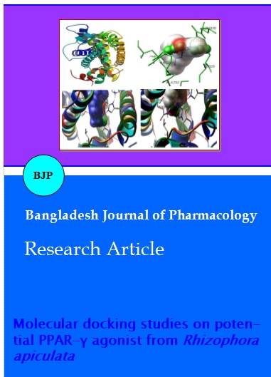
The active site residues of the modelled protein obtained using PBD Sum are HIS323, CYS285, BRL503, MET364, LEU330, GLY284, TYR327, HIS449, LEU453, SER289, TYR473, PHE282, LEU469, TYR327 and ILE 326. Molecular docking studies were performed for modeled PPAR-γ protein with the commercial inhibitor Thiazolidinediones and the selected phytocompounds (Table III). The results were analyzed based on the interaction of H-bonds, interacting residues and binding energy. The better interaction was selected by figuring out the minimum binding energy. The predictions results of all phytoligands were analyzed. The results indicated all the five compounds showed interaction with PPAR-γ protein than the commercial drug TDZ (Figure 1). Among the five compounds, 4-butyl pyridine, 1-oxide gave the best interaction with the amino acid residues (HIS323) of PPAR-γ protein with lowest energy level (-5.1 Kcal/mol) on comparison with the drugs and TDZ showed lowest interaction (-4.3 Kcal/mol) with the modelled protein.
Figure 1: Molecular interaction of PPAR-γ with R. apiculata derived ligands. (A) Secondary structure of PPAR-γ; (B) Interaction with acetamide, N-(4-methylphenyl)-; (C) Interaction with 4-butyl pyridine, 1-oxide; (D) Interaction with clivorine; (E) Interaction with p-aminodiethylaniline; (F) Interaction with thiazolidinediones; Green balls indicates hydrogen bonds
Table III: Molecular interactions of phytocompounds on PPAR-γ
| Ligand | No. of H bonds | Hydrogen bond donor | Length of hydrogen bond(A) | Binding energy | Cluster RMSD | Reference RMSD |
|---|---|---|---|---|---|---|
| Acetamide, N- (4-methylphenyl)- |
2 | ILE326 ARG288 |
2.038 2.116 |
-5.29 | 0.0 | 32.78 |
| 4-Butylpyridine, 1-oxide | 1 | HIS323 | 2.011 | -5.18 | 0.32 | 41.9 |
| Clivorine | 1 | ARG288 | 1.994 | -4.27 | 0.89 | 35.0 |
| p-Aminodiethylaniline | 1 | TYR473 | 2.052 | -4.41 | 0.7 | 42.67 |
| Thiazolidinediones | 4 | TYR473 SER289 HIS449 HIS323 |
2.117 2.101 2.116 1.835 |
-4.28 | 0.21 | 42.73 |
| N-1-Adamantyl-p-methylbenzalimine | No interaction | |||||
The present study concluded that four phytocompounds could be the agonist potential to activate PPAR-γ. The phytocompounds of R. apiculata indicated a better docking simulation and interaction than the existing commercial drug thiazolidinediones.
Table IV: TOPKAT toxicity level
| Compound ID | TOPKAT rat male NTP probability | TOPKAT rat male NTP enrichment | WOE probability | WOE enrichment | WOR score | Rat oral LD50 (g/kg body weight) |
Rat inhalational LC50 (mg/m3/h) |
|---|---|---|---|---|---|---|---|
| CD10097348 | 0.5 | 0.9 | 0.3 | 0.6 | -7.4 | 7.4 | 4.0 |
| CD10374166 | 0.5 | 0.9 | 0.5 | 1.0 | -0.4 | 0.7 | 5.2 |
| CD11106591 | 0.2 | 0.4 | 0.4 | 0.8 | -4.1 | 5.3 | 4.5 |
| CD75952907 | 0.3 | 0.6 | 0.3 | 0.6 | -6.6 | 3.6 | 9.9 |
| CD76675709 | 0.4 | 0.8 | 0.5 | 0.9 | -1.8 | 0.8 | 2.6 |
| CD96875226 | 0.4 | 0.7 | 0.4 | 0.7 | -5.3 | 17.9 | 23.7 |
Acknowledgements
The authors are grateful to the authorities of Annamalai University, Parangipettai, Tamil Nadu and University Grants Commission, New Delhi, India for providing all support during the study period.
References
Braissant O, Foufellie F, Scotto C, Dauca M, Wahli W. Differential expression of peroxisome proliferator-activated receptors (PPARs): Tissue distribution of PPAR-α, -β and -γ in the adult rat. Endocrinology 1996; 137: 354–66.
Evans RM, Barish GD, Wang YX. PPARs and the complex journey to obesity. Nat Med. 2004; 10: 355–61.
Gurudeeban S. Studies on type II diabetes and formulation of tablets from DNA barcoded Rhizophora apiculata Blume derived glycosin on streptozotocin-induced diabetic rats: in vivo, in silico and molecular approaches. Annamalai University, Tamil Nadu, India, 2013, p 283.
Gurudeeban S, Ramanathan T, Satyavani K. Antimicrobial and Radical Scavenging Effect of alkaloid elutes from Rhizophora mucronata. Pharm Chem J. 2013; 47: 50–54.
Jain S, Pulikuri S, Zhu Y, Qi C, Kanwar YS, Yeldandi AV, Rao MS, Reddy JK. Differential expression of the peroxisome proliferator-activated receptor g (PPAR g) and its coactivators steroid receptor coactivator-1 and PPAR-binding protein PBP in the brown fat, urinary bladder, colon and breast of the mouse. Am J Pathol. 1998; 153: 349–54.
James S, Denise EB. Quantification of major tobacco alkaloids by high performance liquid chromatography. J Chromatogr. 1981; 205: 147-54.
Kliewer SA, Willson TM. The nuclear receptor PPAR-g: Bigger than fat. Curr Opin Genet Dev. 1998; 8: 576–81.
Muller G. Let’s shift lipid burden--from large to small adipocytes. Eur J Pharmacol. 2011; 656: 1–4.
Penumetcha M, Santanam N. Nutraceuticals as ligands of PPARγ. PPAR Res. 2012; 97: 1182–86.
Prabhu VV, Guruvayoorappan C. Anti-inflammatory and anti-tumour activity of the marine mangrove Rhizophora apiculata. J Immuotoxicol. 2012; 9: 341–52.
Satyavani K, Gurudeeban S, Ramanathan T, Balasubramanian T. Radical scavenging effect and GCMS identification of alkaloid fractions from Excoecaria agallocha L. Inventi Rapid: Ethnopharmacol. 2014; 1: 1–4.
Schneider M. An oxidized fat containing diet decreases weight gain but increases adiposity in mice fed a low fat diet. Department of Nutrition, Georgia State University, Atlanta, Georgia, 2009, pp 27.
Scholtysik G, Lauener H, Eichenberger H, Burki H, Salzmann R, Muller-Schweinitzer E, Waite R. Pharmacological actions of the antihypertensive agent N-amidino-2-(2,6-dichloro-phenyl) acetamide hydrochloride (BS 100-141). Arzneimittelforschung 1975; 25: 1483-91.
Schoonjans K, Staels B, Auwerx J. The peroxisome proliferator-activated receptors (PPARs) and their effects on lipid metabolism and adipocyte differentiation. Biochem Biophys Acta. 1996; 1302: 93–109.
Spiegelman BM. PPAR-g: Adipogenic regulator and thiazolidinedione receptor. Diabetes 1998; 47: 507–14.
Vinod Prabhu V, Abel Kuruvilla C, Guruvayoorappan C. Potentiating effect of 1, 2-diazole a plant alkaloid on carrageenan and formalin induced paw edema in experimental mice. Int J Pharmcol Pharm Sci. 2012; 4: 3.
Zieleniak A, Wojcik M, Wozniak LA. Structure and physiological functions of the human peroxisome proliferator-activated receptor gamma. Arch Immunol Ther Exp. 2008; 56: 331–45.