Hepatoprotective activity of Chenopodium murale in carbon tetrachloride-induced hepatic damage in rabbits
Abstract
The main purpose of this study was to determine the in vivo hepatoprotective activity of the ethanol extract of whole plant extract of Chenopodium murale (500 and 750 mg/kg; orally) in carbon tetrachloride-induced (0.75 mL/kg; subcutaneously) hepatotoxic rabbit. Silymarin (100 mg/kg/day orally) was used as a standard drug. Hepatotoxic rabbits boosted levels of serum glutamic oxaloacetic transaminase, serum glutamic pyruvic transaminase (SGPT), alkaline phosphatase and total bilirubin. Extracts of C. murale (both doses) proved to have hepatoprotective activity by reducing the elevated level of enzymes. Extract at a dose of 500 mg/kg revealed highly significant (p<0.001) results as compared to 750 mg/kg. Histopathological study of liver tissues additionally authenticated these findings. On the basis of our findings it is concluded that extracts of C. murale can be used in liver disorders.
Introduction
Liver is vital organ, which plays an essential role to regulate many physiological processes in the body. It has significant functions like; metabolism, secretion and storage. It has excessive capacity for the detoxification of toxic constituents and synthesizes beneficial components. Therefore, damage to the liver imposed by hepatotoxic agents is of severe consequences (Shahani, 1999; Subramoniam and Pushpangadan, 1999). Liver diseases are primarily due to toxic chemicals, excess utilization of alcohol, infections and autoimmune diseases. Most of the hepatotoxic chemicals damage liver cells mostly by causing lipid peroxidation and other oxidative damages resulting in generation of highly toxic reactive oxygen species (Recknagel, 1983; Wendel et al., 1987; Dianzani et al., 1991).
Enormous synthesis of reactive oxygen species plays a vital role in the pathogenesis and progression of several diseases involving various organs (Visioli et al., 2000). Carbon tetrachloride is biotransformed into reactive oxidative metabolites by liver microsomal enzymatic system, which results hepatotoxicity (Brent and Rumack, 1993).
Instead of remarkabledevelopments in modern medicine no effective drugs exist, which encourage liver functions and gives protection to the liver from the damage or aids to regenerate hepatocytes (Chattopadhyay, 2003). Due to lack of effective liver-protective drugs in modern medicine, hugenumber of medicinal preparations are endorsed for the treatment of liver disorders (Chatterjee, 2000) and they quite often showed significant relief. Efforts are being made internationally throughout the globe to acquire scientific proofs for these conventionally reported herbal drugs. In the present study, the carbon tetrachloride-induced acute models have been used to assess hepatoprotective activity of C. murale which is used conventionally for prevention of liver disorders.
Chenopodium murale is a member of Chenopodiaceae which is also called "goose foot" family (Marie, 1965). It basically belongs to African and European continents. In later investigations, it was concluded that it is native and abundantly found in subcontinent, so was called "Naturalized". It usually grows rapidly. It is straight upright annual herb usually found in common rough areas like near foot paths of roads and gardens (Khurroet al., 2007). It has been investigated that it is usually found in two types of areas i.e. soily and climatic where temperature normally ranges from 4-45°C (Elkreemiet al., 2009). From medicinal point of view, it is of great value (Ahmad et al., 2003). It grows in flora of Tahat-e-Nasrathi, Tehsil of Karakand in PatolKhel, Bannu, KPK (Khyber Pakhtunkhwa), Pakistan (Khan et al., 2011; Muhammad et al., 2011). It is an evergreen herb and can be found throughout the year (Khan et al., 2011). Also, it is found in Nowshera and Azad Jammu-o-Kashmir with the name of Laalbathua (Singhet al., 2010). It has several medicinal properties reported like: antibacterial, antifungal, insecticidal, cytotoxic, anthelmintic, anti-diaphoretic, stomachic, antispasmodic, emmenagogue, anti-asthmatic, abortifaciant, migraine, digestive problems, sterility, hair loss, anxiolytic, antidepressant and antihypertensive (Ahmad et al., 2003). Its leaves decoction is used in the treatment of jaundice (Jan et al., 2009).
In today practice numbers of herbal formulations are used for treating liver disorders, like Capparis spinosa, Daccus carota, Euphorbia antisyphilitica (Shirwaiker et al., 1996; Bhayee et al., 1995; Saraf et al., 1996). The present pharmacological investigation on aqueous ethanolic extract of different doses of C. murale is conducted for the determination of its hepatoprotective activity against carbon tetrachloride-induced liver damage.
Materials and Methods
Preparation of extract
C. murale whole plant was collected from peripheries of Bahawalpur. Plant was recognized and validated by a texanomist Prof. Tayeb Qureshi. The voucher specimen (Collection No. CM-WB-05-12-051 ) were kept in the herbarium of Department of Pharmacology, Faculty of Pharmacy and Alternative Medicine, the Islamia University of Bahawalpur Pakistan for future reference. 1.5 kg of powdered material of the extract was soaked in 4.0 L of 70%aqueous alcohol. Mixture was shaken occasionally with glass rod for 12 days. On each fourth day of shaking, straining was carried out by sterilized muslin cloth. Menstrum was filtered with filter paper and residue left i.e. marc was again soaked and same process was repeated. The filtrate from each straining was stored in air tight container at 4°C in refrigerator to avoid its deterioration. Finally, whole filtrate was again filtered. Collected filtrate was dried under reduced pressure on rotary evaporator (Laborata 4000 Heidolph, Japan) at 40°C till dryness. 130 g of crude extract was obtained. Percentage yield of dried extract was calculated as 8.66%. Extract was stored in air tight container at 4°C till its experimental evaluation.
Experimental animals
Healthy adult local breed both male and female rabbits were used (each weighing about 1.0-1.5 kg). Animals were held in cages at room temperature (23 ± 12°C). Ten rabbits/cage were accommodated in animal house at Faculty of Pharmacy and Alternative Medicine, the Islamia University of Bahawalpur. Humidity was maintained as 55 ± 15%. Animals were fed properly according to a fixed time table (8 am, 3 pm and 10 pm) with green fodder (Medicago sativa) and tap water was allowed ad libitum. Animals were acclimatized for one week before commencement of experimental studies. Animals were treated according to the guidelines and instructions provided by ethical committee of the Faculty of Pharmacy and Alternative Medicine. Six groups of animals were used each contained ten animals. Group I served as control, Group II received the carbon tetrachloride, Group III received the standard drug silymarin 100 mg/kg (p.o.). The remaining groups received the different amount of extracts (500 mg/kg and 750 mg/kg p.o.) and the hepatotoxin.
Acute toxicity studies
The method adapted from Litchfield and Wilcoxon was used for acute toxicity studies. Male and female mice of either sex were used. Each was weighing 35 ± 5.6 g. During the test, mice were put in the laboratory conditions: standardized boxes, food made of granules, water ad libitum, natural light and ambient temperature at 25-30°C.
Seven groups were made each including six mice. Extract in doses of 1 g/kg, 2 g/kg, 4 g/kg, 8 g/kg, 10 g/kg and 12 g/kg body weight were administered and one untreated control group that received only normal saline. Animals of each group were receiving a specific dose of the extract to be tested and normal saline by oral route using feeding tube. All mice were observed for mortality for a total time period of 72 hours. The dose at which animals died was considered to be the toxic dose (Liechtfield and Wilcoxon, 1949). Animals were died at 8 g/kg.
Hepatoprotective activity of ethanolic extract of C. murale in carbon tetrachloride-induced toxicity
Induction of liver toxicity was carried out by subcutaneous administration of carbon tetrachloride (suspended in olive oil at 1:1) at dose of 0.75 mL/kg body weight. Animals were divided into five groups. Each group consists of 10 rabbits. Total study period was eight days. Carbon tetrachloride was administered 30 min after extract administration on the 7th day of total 8 days study period to all groups except Group I which served as normal control and it received only normal saline. Group II-V received the following treatments from 1st to 7th day of the study (Ahmad and Eram, 2011).
Group I: Normal saline 5 mL/kg; Group II: Negative control (carbon tetrachloride 0.75 mL/kg/day and normal saline at 5 mL/kg/day); Group III: Standard control (silymarin 100 mg/kg/day); Group IV: Extract (500 mg/kg/day); Group V: Extract (750 mg/kg/day).
Assessment of liver function
After 24 hours administration of carbon tetrachloride, rabbits were anaesthetized by ketamine and collection of blood samples (3 mL) was carried out particularly by heart puncture technique (Wright, 1973). Blood samples were allowed to stand at room temperature for coagulation (usually for 45 min) in sterile disposable centrifuge tubes. For obtaining serum, the coagulated blood samples were centrifuged at 2,500 rpm for 15 min. A clear supernatant layer was collected (called serum) from the tubes with the help of micropipette, added into eppendorf tubes and was kept in refrigerator for biochemical parameters like SGOT, SGPT, alkaline phosphatase and total bilirubin (Ahmad and Eram, 2011).
Histopathological studies
For histopathological study, the livers were quickly removed and washed with normal saline and fixed in 10%formalin. Liver sections were made by microtome, dehydrated in ethyl alcohol and cleaned with xylene. Finally these were embedded in paraffin wax. The sections of about 56 mm were cut and paraffin was removed from the embedded section with xylene. Sections were rehydrated with ethyl alcohol and dehydrated with 0.9%NaCl solution. Sections were stained with hematoxylin and Eosin dye. Eventually, these were mounted by Canada balsam. These were then used for the histopathology studies of liver cells architecture under microscope. Their microphotographs were taken (Humason, 1979).
Statistical analysis
The results were presented as mean ± SEM. Unpaired t-test was applied by using Graph Pad Prism software. Differences were significant when p<0.05 (Woodson, 1987).
Results
The in vivo and in vitro hepatoprotective effects of various medicinal plants are generally determined by utilization of acute hepatoxicity model i.e. carbon tetrachloride-induced (Weber et al., 2003). Due to subcutaneous administration of carbon tetrachloride (0.75 mL/kg), there was considerable elevation in the enzyme i.e. SGOT, SGPT and alkaline phosphatase. Total bilirubin level was also raised when compared with normal control. The hepatoprotective activity of extracts on acute hepatotoxicity model induced by carbon tetrachloride has been outlined in Table I.
Table I: Effects of ethanolic extract of C. murale (whole plant) on rabbits serum biochemical parameters after carbon tetrachloride administration
| Groups | SGPT (IU/L) |
SGOT (IU/L) |
Alkaline phosphatase (IU/L) |
Total bilirubin (mg/dL) |
Liver damage |
|---|---|---|---|---|---|
| Normal control | 65.3 ± 2.9 | 49.2 ± 1.8 | 218.1 ± 3.7 | 0.7 ± 0.1 | 0 |
| Negative control | 402.4 ± 51.4 | 398.7 ± 6.6 | 371.5 ± 4.6 | 1.4 ± 0.0 | 3 |
| Standard control (Silymarin) | 203.3 ± 6.8 | 232.3 ± 3.4 | 249.0 ± 4.0 | 0.8 ± 0.1 | 1 |
| Extract (500 mg/kg + Carbon tetrachloride) | 276.3 ± 3.4 | 256.2 ± 5.2 | 278.8 ± 2.8 | 1.2 ± 0.1 | 1 |
| Extract (750 mg/kg + Carbon tetrachloride) | 217.2 ± 7.0 | 205.7 ± 5.9 | 244.5 ± 5.4 | 0.8 ± 0.1 | 1 |
| Values are represented as Mean ± S.E.M. (n=10). 0 = Normal, +1 = Mild, +2 = Moderate and +3 = Severe. Microphotographs of liver sections of different groups | |||||
The groups treated with C. murale extract exhibited the low levels of SGOT, SGPT, alkaline phosphatase and total bilirubin when compared with negative control group. C. murale extract at 500 mg/kg and 750 mg/kg doses exhibited hepatoprotection in comparison to standard silymarin (100 mg/kg). C. murale extract at 500 mg/kg dose gave more significant (p<0.05) results in comparison with 750 mg/kg.
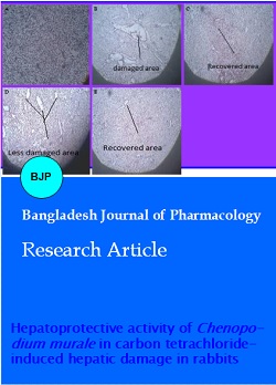
Histopathological studies of livers were conducted after 24 hours of induction of hepatotoxicity. The changes found were necrosis, infiltration of inflammatory cells, fats congregation, number of vacuoles were increased and hepatic stenosis. These events were because of free radical CCl3. (trichloromethyl radical) generated due to carbon tetrachloride metabolism. Histopathological changes are exhibited in Figure 1. C. murale extract at both doses restored disturbed hepatic cell morphology to normal when compared with standard and negative controlled group.
Figure 1: Histopathological slides of liver of different animal groups. Normal control (A); Carbon tetrachloride negative control (B); Standard Control(C); Test group 1(D); Test group 2 (E)
Serum SGOT, SGPT and alkaline phosphatase elevation reflect liver damage due todisintegration of cell membrane and necrosis. This causes cellular leakage and ultimately functional integrity of cell is lost (He and Aoyama, 2003). Also, elevated level of serum total bilirubin directs jaundice (Sturgill and Lambert, 1997). Serum enzyme level and histopathological findings direct the intensity and kind of liver injury. Above mentioned enzymes level was almost restored by C. murale extract showing hepatoprotective activity.
Discussion
Cytochrome i.e. CYP 2E1, CYP 2B1 or CYP 2B2 and possibly CYP 3A activate carbon tetrachloride to form CCl3* (trichloromethyl radical) which is a free radical. Free radicals induces oxidative stress that leads to cell membrane injury i.e. plasma membrane disintegration which result in alteration in metabolic processes. Reactive oxygen species play an important role in the pathogenesis of various degenerative diseases and have been found to implicate in atherosclerosis, liver disorders, lung and kidney damage, aging and diabetes mellitus. In liver disorders, the ability of natural antioxidant system is impaired (Johnston and Korening, 1998). Reactive oxygen species is constantly removed from the cell as it is produced, however, when its concentration is beyond the limits it results in deleterious effects on cell. Eventually peroxidation and alkylation take place (Halliwel and Gutteridge, 1990), resulting in destruction of basic skeleton of the cell, alteration in the functional capability of mitochondria and disturbance in the homeostasis of ions (Brattinet al.,1984).
The activities of serum marker enzymes like SGOT, SGPT and alkaline phosphatase can make assessment of liver function. When liver cell membrane is damaged, these enzymes normally located in the cytosol are released in to blood stream. Their estimation in the serum is a useful quantitative marker of the extent and type of hepatocellular damage (Sallie, 1999). In common, SGOT and alkaline phosphatase are present in high amount in liver due to hepatic necrosis (Shah et al.,2002). Bilirubin is a break down product of heme and present in the cytoplasm of hepatic cells. Whenever there is damage to the liver cells, it leaks into blood stream which authenticates liver damage (Nkosi et al.,2005).
In the current study, enzymes level (SGOT, SGPT and alkaline phosphatase) was elevated considerably in negative control group with damaged liver morphology. The extract at both doses (500 mg/kg and 750 mg/kg) reduced the elevated level of enzymes and restored the normal morphology. This alleviation of enzymes and restoration of damaged liver cell morphology to normal is because of decrease in lipid peroxidation, aggravated by free radicals i.e. CCl3. (trichloromethyl radical), CCl3OO• (trichloromethylperoxy radical) (Weber et al., 2003). The decrease in enzymes level is due to the presence of antioxidants in C. murale which has free radical scavenging activity. This reflects the protection of structural and functional integrity of carbon tetrachloride damaged liver cells. Also, restoration of serum alkaline phosphatase and total bilirubin level nearer to normal shows consistency of biliary functions. Phytochemical analysis shows that C. murale contains; volatile oils, saponines, gerniol, flavonoids, alkaloids, sterols, and coumarins (Ahmad et al., 2011). Flavonoids and saponins are known to hold hepatoprotective action in animals (Tran et al., 2001).
Flavonoids block lipid peroxidation in cell membranes due to their high antioxidant activity and free radical scavenging property (Halliwel and Guitreg, 1990). Kaempferol is a reported flavonoid found in C. murale and has strong antioxidant activity which helps to prevent oxidative harm to cells, lipids and DNA. It also contains beta-setosterol which is a reported hepatoprotective agent. (Ahmad et al., 2011). Coumarins in C. murale have potent antioxidant effect and hence strong hepatoprotective activity (Amacaet al., 2011).
In the current study, extract caused a substantial inhibition in SGOT, SGPT and alkaline phosphatase activities towards the respective normal range with contemporary reduction of raised bilirubin level (Mukherjee, 2002). This shows that extract restored the structural integrity of the hepatocellular membrane damaged by carbon tetrachloride which was authenticated by histopathological investigation. The occurrence of saponins, flavonoids and tannins in extract may participate the hepatoprotective activity. The hepatoprotective effect of the extract of C. murale may be because of its capability to halt the bioactivation of carbon tetrachloride and its powerful antioxidant potential by scavenging the free radicals.
Conclusion
The aqueous ethanolic extract of C. murale has potent hepatoprotective action upon carbon tetrachloride-induced hepatic damage in rabbit.
Ethical Issue
Animals were treated according to the guidelines and instructions provided by ethical committee of the Faculty of Pharmacy and Alternative Medicine.
Acknowledgement
The authors are thankful to Dr. Naveed Akhtar, Chairman, Department of Pharmacy, Faculty of Pharmacy and Alternative Medicine the Islamia University of Bahawalpur for the provision of all the required facilities in accomplishing this project.
References
Ahmad B, Jan Q, Choudhary MI, Nisar M. Phytochemical evaluation of Chenopodium murale L. Asian J Plant Sci. 2003; 2: 1072–78.
Ahmad M, Eram S. Hepatoprotective studies on Haloxylon salicornicum: A plant from cholistan desert. Pakistan J Pharm Sci. 2011; 24: 377-82.
Amaca M, Bilgin HM, Obay BD, Diken H, Kelle M, Kale E. The hepatoprotective effect of coumarin and coumarin derivates on carbon tetrachloride-induced hepatic injury by antioxidant activities in rats. J Physiol Biochem. 2011; 67: 569-76.
Brattin WJ, Pencil SD, Waller RL, Glende EA, Reckenjel O. Assessment of role of calcium ion in hydrocarbon hepatotoxicity. Environ Health Perspect. 1984; 57: 321-23.
Brent JA, Rumack BH. Role of free radicals in toxic hepatic injury. II. Are free radicals the cause of toxin-induced liver injury? J Toxicol Clin Toxicol. 1993; 31: 173-96.
Bhayee A, Sarkar A, Chatterjee M. Hepatoprotective activity of carrot (Dacuscarota Linn) against CCl4-intoxication in mouse liver. J Ethnopharmacol. 1995; 47: 69-74.
Chattopadhyay, RR. Possible mechanism of hepatoprotective activity of Azadirachta indica leaf extract. Part II. J Ethnopharmacol. 2003; 89: 217-19.
Chatterjee TK. Medicinal plants with hepatoprotective properties in herbal opinions. vol. III. Calcutta, Books and Allied (P) Ltd., 2000, p 135.
Dianzani, MU, Muzia G, Biocca ME, Canuto RA. Lipid peroxidation in fatty liver induced by caffeine in rats. Int J Tissue React. 1991; 13: 79-85.
Elkreemi A, Eideh RA, Zaiter A. The growth of Chenopodium murale irrigated with polluted and unpolluted water: A modeling approach. Australian J Basic Appl Sci. 2009; 3: 1827-37.
Halliwel B, Gutteridge JM. Role of free radicals and catalytic metal ions in human disease: An overview. Methods in enzymology. 1990; 186: 1-85.
Handa SS, Sharma A. Hepatoprotective activity of andrographolide from Andrographis paniculata against carbon tetrachloride. Indian J Med Res. 1990; 92: 276-83.
He G, Aoyama Y. Effects of adding some dietary fibers to a cystine diet on the activities of liver antioxidant enzymes and serum enzymes in rats. Biosci Biotechnol Biochem. 2005; 67: 617-21.
Humason GL. Animal tissue techniques. 4th ed. San Francisco, WH Freeman and Company, 1979, pp 1-16.
Johnston DE, Korening C. Mechanism of early carbon tetrachloride toxicity in cultured rat hepatocytes. Pharmacol Toxicol. 1998; 83: 231-39.
Jan G, Khan MA, Jan F. Medicinal value of the Asteraceae of DirKohistan Valley, NWFP. Ethnobotanical Leaflets. 2009; 13: 1205.
Khan M, Farrukh H, Shahana M. Preliminary floristic range of Tehsil Takht-e-Nasrati Pakistan. Int J Biosci. 2011; 23: 88-99.
Khurro AA, Dar GH, Khan ZS, Malik AH. Exploring an inherent interface between taxonomy and biodiversity: Current problems and future challenges. J Nat Conserv. 2007; 15: 256-61.
Liechtfield CJT, Wilcoxon F. A simplified method of evaluating dose-effect experiments. J Pharmacol Exp Ther. 1949; 96: 99-113.
Marie CN. In: Gardens of Hawaii. Bishop Museum Press, 1965, p 331.
Muhammad Z, Wazir SM, Farooq A, Hussain Z. Distribution and checklist of weeds in maize crop of frontier region Bannu, Khyber Patunkhwa, Pakistan. Pakistan J Weed Sci. 2011; 17: 373-79.
Mukherjee PK. Quality control of herbal drugs, 1st ed. New Delhi, Business Horizons Pharmaceutical Publication, 2002, p 531.
Nkosi CZ, Opoku AR, Terblanche SE. Effect of pumpkin seed (Cucurbita pepo) protein isolate on the activity levels of certain plasma enzymes in CCl4-induced liver injury in low protein fed rats. Phy ther Res. 2005; 19: 341–45.
Recknagel RO. A new direction in the study of carbon tetrachloride hepatotoxicity. Life Sci. 1983; 33: 401-08.
Shahani S. Evaluation of hepatoprotective efficacy of APCL: A polyherbal formulation in vivo in rats. Indian Drugs. 1999; 36: 628-31.
Subramoniam A, Pushpangadan P. Development of phytomedicine for liver diseases. Indian J Pharmacol. 1999; 31: 166-75.
Saraf S, Dixit VK, Patnaik GK, Tripathi SC. Antihepatotoxic activity of Euphorbia antisyphilitica. Indian J Pharm Sci. 1996; 58: 137-41.
Shirwaiker A, Sreenivasan KK, Krishnanand BR, Vasanth Kumar A. Chemical investigation and antihepatotixic activity of the root bark of Capparis spinosa. Fitoterapia 1996; 67: 200-04.
Singh T, Sharma A, Sharma LR, Amandeep S. Common weeds of rabi (winter) crops of tehsil Nowshera, District Rajouri (Jammu & Kashmir), India. Pakistan J Weed Sci. 2010; 16: 39-45.
Sturgill MG, Lambert GH. Xenobiotic-induced hepatotoxicity: Mechanisms of liver injury and methods of monitoring hepatic function. Clin Chem. 1997; 43: 1512-26.
Shah M, Patel P, Phadke M, Menon S, Sane RT. Evaluation of the effect of aqueous extract from powders of root, stem, leaves and whole plant of Phyllanthus debilis against CCL4 induced rat liver dysfunction. Indian Drugs. 2002; 39: 333-37.
Sallie R, Tredger JM, William R. Drugs and the liver. Biopharm Drug Dispos. 1999; 12: 251-59.
Tran QL, Adnyana IK, Tezuka Y, Nagaoka T, Tran QK, Kadota S. Triterpene saponins from Vietnamese ginseng (Panax vietnamensis) and their hepatocytoprotective activity. J Nat Prod. 2001; 64: 456-61.
Visioli F, Keaney JF, Halliwell B. Antioxidant and cardiovascular disease; panaceas or tonics for tired sheep? Cardivasc Res. 2000; 47: 409.
Weber LW, Boll M, Stampfl A. Hepatotoxicity and mechanism of action of haloalkanes: Carbon tetrachloride as a toxicological model. Crit Rev Toxicol. 2003; 33: 105-36.
Woodson RR. Statistical methods for the analysis of biochemical data, series in probability and mathematical statistics, New York, Wiley, 1987, p 316.
Wright JW. The effects of a heart puncture blood sample upon body water maintenance in rats. Physiol Behavior. 1973; 10: 407-10.
Wendel A, Feurensteins S, Konz KH. Acute paracetamol intoxication of starved mice leads to lipid peroxidation in vivo. Biochem Pharmacol. 1987; 28: 2051-53.