Anti-diabetic potential of Conocarpus lancifolius
Abstract
The anti-diabetic activity of Conocarpus lancifolius extract was investigated in vitro, as alpha glucosidase inhibition and in vivo as alloxan induced diabetic rabbits with other biochemical parameters (LDL, HDL, SGPT, SGOT, cretinine, urea and triglyceride). Alpha-glucosidase inhibition activity was performed by using acorbose as standred. Methanolic extract show alpha-glucosidase inhibition activity. The dose of 200 mg/kg body weight significantly (p<0.05) decreases the blood glucose level, plasma total cholesterol, triglycerides and LDL in treated rabbits as compared to diabetic rabbits. This dose significantly increased the level of HDL in treated group. The activity of SGOT and SGPT also significantly (p<0.05) decreased in treated diabetic rabbits. Phytochemical studies show the presence of glycosides, tannins, saponins and terpenoids. The anti-diabetic potential is may be due to its saponin contents.
Introduction
World Health Organization estimated that for health care needs, four-fifth of the total populations till confidence on the plant medicine (Farnsworth et al., 1985). Among the plants, Carica papaya (Sadeque and Begum, 2010), Carissa spinarum (Hegde and Joshi, 2010), Cestrum nocturnum (Qadir et al., 2014), Chenopodium murale (Saleem et al., 2014), Cocculus hirsutus (Thakare et al., 2009), Convolvulus arvensis (Ali et al., 2013), Dodonaea viscosa (Khan et al., 2013), Khamira Gaozaban Ambri Jadwar Ood Saleeb Wala (Akhtar et al., 2013), OfIpomoea staphylina (Bag and Mumtaz, 2013), Suaeda fruticosa (Rehman et al., 2013), Trianthema decandra (Balamurugan and Muthusamy, 2008) and Trichodesma sedgwickianum (Saboo et al., 2013) showed hepatoprotective effect.
Conocarpus lancifolius Engl., an ornamental tree, is native to coastal and riverine areas of East Africa (Baroon et al., 2012). The leaves are glossy in appearance with relatively fewer trichomes on both surfaces (Redha et al., 2011). The anti-diabetic capacities of C. lancifolius consumed locally in Pakistan have not been presented. However, up to now a little phytochemical or pharmacological work was done.
Materials and Methods
Collection and extraction of plant material
The plant material was collected from surroundings of Lahore (Pakistan). The plant was identified as C. lancifolius Engl. by Prof. Altaf Ahmad Dasti, Taxanomist Institute of pure and applied Biology, Bahauddin Zakariya University, Multan.
For the purpose of effective extraction, whole plant of the C. lancifolius was dried under the shade for 17 days. The plant material was coarse grounded and then grounded to fine powder. This powder was subjected to extraction through the simple maceration method at room temperature occasionally shaking for 24 hours. The weighted amount (1 kg) of plant material was kept in large size well caped containers and added 1200 mL of methanol as solvent, to get the maximum extraction results; the flask having mixture was placed in ultrasonic bath for about half hour daily. Filtration was done after the addition of solvent. Same process was carried out 3 times to get the maximum extracted volume. The methanol (18.3 g) extract was collected in separate sample bottles and designated with code as CLM.
Phytochemical screening
The presence of alkaloids, glycosides, tannins, flavonoids, anthraquinones and saponins were screened according to the method of (Trease and Evan, 1998).
Alkaloids: About 5 g of powdered drug was boiled in dilute hydrochloric acid. It was filtered and made alkaline by the addition of dilute ammonia solution. This alkalinized solution was extracted with 5 mL of chloroform. The chloroform layer was then extracted with 10 mL dilute acetic acid. To the acetic acid extract, few drops of Dragendorff’s reagent was added (Orange precipitate or turbidity indicate the presence of alkaloids).
Braemer’s test for tannins: 5 g of drug was extracted with methanol. To methanolic extract, 10% alcoholic ferric chloride solution was added. Dark blue or greenish grey coloration of the solution indicates the presence of tannins in the drug.
Saponins: 5 g of powdered drug was vigorously shaken with water and noted for production of froth (persistent froth for 20 min indicates the presence of saponins).
Borntrager’s test for free anthraquinones: 5 g of powdered drug was extracted with hot water and filtered while hot. On cooling, it was extracted with carbon tetrachloride. The carbon tetrachloride was separated and washed with water and shaken with dilute ammonia solution (pink to cherry-red color in ammonia layer indicate the presence of free anthrax-quinones).
Modified Borntrager’s test for bound anthraquinones: 5 g of powdered drug was extracted with ferric chloride solution and hydrochloric acid. It was heated on water bath for 10 min and filtered while hot. On cooling, it was extracted with carbon tetrachloride. The carbon tetrachloride was separated and washed with water and shaken with dilute ammonia solution color (intense pink to cherry-red coloration of ammonia layer indicate the presence of anthraquinones glycosides).
Shinoda test for flavonoids: 5 g of drug was extracted with methanol. To methanol extract, a piece of magnesium ribbon and 1 ml of concentrated hydrochloric acid were added. (Pink red or red coloration of the solution indicate the presence of flavonoids in the drug).
In vitro α-glucosidase inhibition assay
The α-glucosidase inhibitory activity was assessed by the standard method (Dong et al., 2012) with slight modifications. Briefly, a volume of 60 μL of sample solution and 50 μL of 0.1 M phosphate buffer (pH 6.8) containing α-glucosidase solution (0.2 U/mL) was incubated in 96 well plates at 37 ºC for 20 min. After pre-incubation, 50 μL of 5 mM p-nitrophenyl-α-D-glucopyranoside (PNPG) solution in 0.1 M phosphate buffer (pH 6.8) was added to each well and incubated at 37ºC for another 20 min. Then the reaction was stopped by adding 160 μL of 0.2 M NaCO3 into each well, and absorbance readings (A) were recorded at 405 nm by microplate reader and compared to a control which had 60 μL of buffer solution in place of the extract. For blank incubation (to allow for absorbance produced by the extract), enzyme solution was replaced by buffer solution and absorbance recorded. The concentrations of test compounds which inhibited the hydrolysis of substrates (butyrylthiocholine) by 50% (IC50) were determined by monitoring the effect of increasing concentrations of these compounds in the assays on the inhibition values (Rehman et al., 2013). The IC50 values were then calculated using the EZ-Fit Enzyme Kinetics program (Perrella Scientific Inc., Amherst, USA).
In vivo anti-diabetic study
Animal selection and treatment
Thirty male rabbits each of weighing 1-1.5 kg were used and all rabbits were checked for evidence of any infections. All the rabbits were housed in the animal house of the Department of Pharmacy B.Z.U Multan. They were housed in the steel cages under standard laboratory condition (light period 8:00 am to 8:00 pm 21± 2°C, relative humidity 55%, green fodder and water was available as labatum). The protocol was approved by Ethical Committee of Department of Pharmacy Bahauddin Zakariya University Multan. Groups were control (non-diabetic), control group (received vehicle only, diabetic), alloxan induced diabetic rabbits treated with C. lancifolius 100 mg/kg, alloxan induced diabetic rabbits treated with C. lancifolius 200 mg/kg, and alloxan-induced diabetic rabbits treated with diamicron 80 mg/kg (Hader et al., 1994).
Induction of diabetes
Animals were devided into five experimental groups (control, diabetic, diabetic treated with 100 mg/kg body weight C. lancifolius diabetic treated with 200 mg/kg body weight, C. lancifolius and diabetic treated with diamicron 80 mg/kg) Each group contained six rabbits. Before starting experiment the animals in latter four groups were injected. 150 mg/kg of alloxan (sigma chemical co,) dissolved in 10% isotonic saline to induce diabetes (Issa et al., 2000). The alloxan is selective beta-cells cytotoxic drug. It produced all signs and symptoms of diabetes. i.e. hyperglycemia, glycosuria, polydipsia, polyurea, etc. The control group was injected the same volume of isotonic solution as that of diabetic received. The blood glucose level of surviving rabbits was checked after three days of Alloxan injection and rabbits with blood glucose level more than 250 mg/dL on 12 hours fasting were considered as diabetics (Fridewald et al., 1979).
The last two groups were given the C. lancifolius extract, i.e. Group 1 received 100 mg/kg C. lancifolius extract daily for 30 days; Group 2 received 200 mg/kg C. lancifolius extract daily for 30 days.
Biochemical analysis
The plasma glucose levels were measured, using a glucose oxidase kit (Accu Check Advantage Rosche Chemicals, Switzerland). The plasma total cholesterol, triglyceride, HDL, and ALT & AST levels were evaluated by enzymatic test kits (Randox). LDL level was calculated by using following formula:
LDL = Total cholesterol – HDL - (triglyceride/5)
Creatinin clearance was determined with the help of Chemistry analyzer Micro Lab 200 (Merck).
Statistical analysis
The experimental results were expressed as mean ± standard deviation (SD) of three replicates. The data were subjected to one-way analysis of variance (ANOVA) Turkey’s Test was used to test for differences among means for which ANOVA indicated a significant.
Result and Discussion
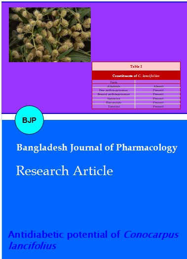
The phytochemical screening of arial part of C. lancifolius is shown in Table I. In vitro α-glucosidase inhibitory activities of the plant extracts aforementioned are summarized in Table II. Methanol extract possessed excellent α-glucosidase inhibitory activity even at low concentration. Methanol extract studied showed the most potent inhibitory activity 99.8% with IC50 found to be 14.9 mg/mL compared to the standard (acorbose) which exhibited 92.2% inhibition with IC50 found to be 14.9 mg/mL inhibition on α-glucosidase. Table III shows the antidiabetic effect of C. lancifolius treated (100 and 200 mg/kg) as compared to treated with 80 mg/kg diamicron, control and diabetic. Blood glucose concentration of diabetic rabbits 269.2 ± 5.8 mg/dL (mean ± SD) was increased significantly (p≤0.05) as compared to the control group. In control group level of blood glucose was 93.2 ± 5.4 mg/dL. Treatment with methanolic extract 200 mg/kg of C. lancifolius significantly decreased the blood glucose levels of diabetic treated group with mean value 136.0 ± 3.15 mg/dL (p<0.05) and their glucose levels were within normal range (75-150 mg/dL). Diamicron has no significant effect (226.2 ± 4.1 mg/dL) as compared with C. lancifolius methanol extract (136 ± 3.1 mg/dL).
Table I: Constituents of C. lancifolius
| Tests | |
|---|---|
| Alkaloids | Absent |
| Free anthraquinones | Present |
| Bound anthraquinones | Present |
| Saponins | Present |
| Flavonoids | Present |
| Tannins | Present |
Table II: Inhibitory effects of C. lancifolius on α-glucosidase
| Concentration (mg/mL) | % Inhibition |
|---|---|
| 0.5 | 99.1 |
| 0.25 | 98.7 |
| 0.125 | 99.8 |
| 0.0625 | 98.9 |
| 0.03125 | 98.1 |
| 0.0156 | 99.1 |
| 0.0078 | 99.0 |
| 0.0039 | 98.5 |
Table III: Effect of C. lancifolius extract on blood glucose level in alloxan-induced diabetic rabbits
| No. of days | Glucose level | ||||
|---|---|---|---|---|---|
| Control | Diabetic | Diabetic with | |||
| Extract (100 mg/kg) | Extract (200 mg/kg) | Diamicran (80 mg/kg) | |||
| 0 | 93.0 (5.1) |
270.4 (3.3) |
270.0 (5.4) |
269.0 (4.7) |
272.0 (5.9) |
| 7 | 91.0 (4.3) |
268.2 (4.1) |
155.0 (5.1) |
144.5 (5.5) |
232.1 (3.2) |
| 14 | 93.0 (5.1) |
271.0 (6.3) |
146.2 (3.7) |
138.2 (5.6) |
230.3 (4.3) |
| 21 | 97.0 (4.3) |
271.0 (4.4) |
134.7 (4.2) |
130.8 (4.4) |
222.4 (5.4) |
| 28 | 92.0 (4.4) |
265.0 (5.6) |
130.3 (5.1) |
124.6 (3.4) |
220.3 (4.4) |
| Number of rabbits in each group =6; Values are expressed as mean and SD within the parenthesis | |||||
Table IV shows the antihyperlipidemic effect of C. lancifolius extract in alloxan induced diabetic rabbits. Hypercholestrolemia and hypertriglyceridemia have been reported to occur in alloxan-induced diabetic rabbits. Total cholesterol, triglyceride and LDL mg/dL values increased significantly (p≤0.05) in Diabetic rabbits. Whereas HDL mg/dL values decreased significantly (p≤0.05) in diabetic rabbits. C. lancifolius extract treatment decreased the cholesterol, triglyceride and LDL values and increased HDL values significantly (p≤0.05) in diabetic treated rabbits. So, C. lancifolius not only decreases the blood glucose level but also eliminate the side effects associated with diabetes in rabbits.
Table V shows the liver function effects of C. lancifolius extract in diabetic rabbits. Because diabetes mellitus is the commonest cause of liver failure and hepatomegaly (Chatila and West, 1996) which itself represents a huge and rapidly increasing problem. Liver and kidney glutathione levels have been reported to decrease in experimental diabetic rats (Loven et al., 1986).
SGOT and SGPT Values increased significantly (p≤0.05) in diabetic rabbits. C. lancifolius extract treatment decreased (p≤0.05) SGOT and SGPT values in diabetic rabbits (Table VI).
Table IV: Effect of C. lancifolius extract on total cholesterol and triglycerides level in alloxan-induced diabetic rabbits
| No of days | Total cholesterol (mg/dL) | Triglyceride (mg/dL) | ||||||||
|---|---|---|---|---|---|---|---|---|---|---|
| Control | Diabetic | Diabetic with | Diabetic | Diabetic with | ||||||
| Extract (100 mg/kg) | Extract (200 mg/kg) | Diamicran (80 mg/kg) | Control | Diabetic | Extract (100 mg/kg) | Extract (200 mg/kg) | Diamicran (80 mg/kg) | |||
| 0 | 41.2 (3.5) |
64 (4.7) |
64.3 (2.9) |
66.1 (3.1) |
66.9 (3.9) |
57.2 (8.3) |
139 (9.1) |
140.4 (11.5) |
135.1 (13.3) |
137.9 (9.3) |
| 7 | 45.3 (2.6) |
66 (5.1) |
54.5 (3.6) |
52.1 (2.7) |
58.3 (4.3) |
58.1 (8.5) |
135.4 (8.5) |
104 (9.3) |
99.3 (12.2) |
104 (9.5) |
| 14 | 43.1 (2.3) |
65.5 (5.5) |
53.4 (3.1) |
52 (3.1) |
55 (3.7) |
57.5 (9.1) |
136.1 (9.8) |
96.5 (10.2) |
99.5 (10.3) |
100 (12.2) |
| 21 | 42 (3.4) |
64.2 (5.2) |
52 (2.3) |
51 (3.5) |
55.1 (3.8) |
57.4 (12) |
136 (7.5) |
74 (8.5) |
80 (8.7) |
93 (13.1) |
| 28 | 43.7 (3.1) |
65 (5.5) |
52.1 (2.3) |
51.3 (3.5) |
54.2 (3.6) |
59 (10.1) |
139 (11) |
74 (9.3) |
74.2 (8.5) |
81.1 (11.5) |
| Number of rabbits in each group = 6; Values are expressed as mean and SD within the parenthesis | ||||||||||
Table V: Effect of C. lancifolius extract on HDL and LDL in alloxan-induced diabetic rabbits
| No of days | HDL (mg/dL) | LDL (mg/dL) | ||||||
|---|---|---|---|---|---|---|---|---|
| Control | Diabetic | Diabetic with | Diabetic with | |||||
| Extract (100 mg/kg) | Extract (200 mg/kg) | Diamicran (80 mg/kg) | Extract (100 mg/kg) | Extract (200 mg/kg) | Diamicran (80 mg/kg) | |||
| 0 | 28.1 (3.5) |
34.2 (2.0) |
35.3 (2.5) |
36.1 (3.1) |
35.1 (2.5) |
8.9 (2.5) |
9.7 (3.1) |
8.9 (3.5) |
| 7 | 28 (3.1) |
35.4 (2.7) |
36 (3.1) |
34.2 (2.7) |
34.5 (3.0) |
7.3 (3.1) |
7 (3.1) |
7.7 (3.2) |
| 14 | 29.4 (3.5) |
34 (2.3) |
34.5 (3.1) |
34 (2.5) |
36.2 (3.2) |
7.1 (2.7) |
7.1 (2.4) |
7.5 (2.6) |
| 21 | 28.2 (2.5) |
34.5 (3.1) |
34.7 (3.0) |
33.7 (2.2) |
34.5 (3.1) |
6.9 (3.5) |
6.7 (3.1) |
7.9 (3.1) |
| 28 | 27.5 (3.1) |
35 (2.8) |
36.2 (3.7) |
36.9 (2.9) |
33.5 (2.7) |
7.2 (3.2) |
7.3 (3.1) |
7.2 (4.1) |
| Number of rabbits in each group =6; Values are expressed as mean and SD within the parenthesis | ||||||||
Table VI: Effect of C. lancifolius extract on serum SGPT and SGOT (IU/L) in alloxan-induced diabetic rabbits
| No. of days | Serum SGPT (IU/L) | Serum SGOT (IU/L) | ||||||
|---|---|---|---|---|---|---|---|---|
| Control | Diabetic | Diabetic with | Diabetic with | |||||
| Extract (100 mg/kg) | Extract (200 mg/kg) | Diamicran (80 mg/kg ) | Extract (100 mg/kg) | Extract (200 mg/kg) | Diamicran (80 mg/kg ) | |||
| 0 | 202 (51.1) |
206 (15.5) |
206 (11.5) |
203.5 (12.1) |
204 (13.5) |
197 (13.5) |
201 (5.1) |
203 (16.4) |
| 7 | 203 (12.5) |
203.5 (12.1) |
188.3 (25.3) |
183.3 (11.5) |
189.3 (15.3) |
172 (5.3) |
176 (36.3) |
180.3 (9.5) |
| 14 | 200 (13.5) |
204 (9.8) |
186 (13.5) |
185.3 (15.1) |
185.3 (5.1) |
160 (3.3) |
172 (12.5) |
178.5 (3.5) |
| 21 | 204 (15.1) |
202 (13.3) |
178 (11.1) |
180 (13.3) |
186 (13.5) |
170 (12.5) |
168 (13.2) |
176.3 (12.1) |
| 28 | 201 (13.5) |
205 (25.1) |
178.9 (15.1) |
180.5 (23.3) |
188.3 (3.5) |
171 (3.2) |
165 (16.1) |
176 (13.5) |
| Number of rabbits in each group=6; Values are expressed as mean and SD within the parenthesis | ||||||||
The effect of C. lancifolius extract in alloxan-induced diabetic rabbits on creatinine clearance. Because chronic diabetes mellitus causes renal failure (Sacks, 1997).
Creatinine clearance mg/dL values were not significantly (p≤0.05) changed in diabetic rabbits.
Blood urea level of diabetic rabbits is not significantly increased and treatment with C. lancifolius has not significant effect on blood urea and creatinine concentration.
Diabetes mellitus is a chronic disorder of metabolism caused by an absolute or relative lack of insulin. It is characterized by hyperglycemia in postprandial and fasting state, and its severe form is accompanied by ketosis and protein wasting (Athanasakis et al., 2010).
Diabetes is one of the world's greatest health problems, affecting about 171 million people and most of these will be dominated by those suffering from type II diabetes (Moller, 2001).
In diabetes mellitus there is heterogencity, so need for appropriate therapy increased. There is a use of traditional medicinal plants to overcome the complications of diabetes (Alamgeer et al., 2013). Our studies offer a natural key to unlock the complications in future. α-glucosidase inhibitor inhibits the disaccharide digestion and impedes the postprandial glucose excursion to enable overall smooth glucose profile (Casirola and Ferraris, 2006). Thus, natural products of great structural diversity are still a good source for searching for such inhibitors, thereby motivating to explore biologically active compounds from the highly diverse plants.
Differences in social structure, psychic stress, obesity, hormonal imbalance and heredity are optimizing the growth of pandemic. At present, the treatment of diabetes mainly involves a sustained reduction in hyperglycemia by the use of biguanides, thiazolidinediones, sulphonylureas, D-phenylalanine derivatives, meglitinides and α-glucosidase inhibitors in addition to insulin. However, due to unwanted side effects the efficacies of these compounds are debatable and there is a demand for new compounds for the treatment of diabetes (Oubre et al., 1997). Hence, plants have been suggested as a rich, as yet unexplored source of potentially useful antidiabetic drugs. However, only a few have been subjected to detailed scientific investigation due to a lack of mechanism-based available in vitro assays (Bell, 1991). These efforts may provide treatment for all and justify the role of novel traditional medicinal plants having antidiabetic potentials. The methanolic extract also prevents body weight loss in diabetic rabbits.
Conclusion
The methanolic extract of C. lancifolius have prominent anti-diabetic potential due to gluconeogenesis suppession in alloxan-induced diabetic rabbits may be due to saponins contents of C. lancifolius.
Acknowledgement
The authors are grateful to the Pharmacy Department, Bahauddin Zakariya University Multan, Pakistan.
Ethical Issue
The protocol was approved by Ethical Committee of Department of Pharmacy Bahauddin Zakariya University Multan.
References
Abdel-Hameed ES, Bazaid SA, Shohayeb MM, El-Sayed MM, El-Wakil. Phytochemical studies and evaluation of antioxidant, anti-cancer and antimicrobial properties of Conocarpus erectus L. growing in Taif, Saudi Arabia. Eur J Med Plants. 2012; 2: 93-112
Abdel-Hassan IA, Abdel-Barry JA, Tariq Mohammeda S. The hypoglycemic and antihyperglycemic effect of citrullus colocynth fruit in normal and diabetic rabbits. J Enthopharmacol. 2000; 71: 325-30.
Akhtar MS, Asjad HMM, Bashir S, Malik A, Khalid R, Gulzar F, Irshad N. Evaluation of antioxidant and hepatoprotective effects of Khamira Gaozaban Ambri Jadwar Ood Saleeb Wala (KGA). Bangladesh J Pharmacol. 2013; 8: 44-48.
Alamgeer, Rashid M, Bashir S, Mushtaq MN, Khan HU, Malik MNH, Qayyum A, Shafeeq ur Rahaman M. Comparative hypoglycemic activity of different extracts of Teucrium stocksianum in diabetic rabbits. Bangladesh J Pharmacol. 2013; 8: 186-93.
Al-hader AA, Hassan ZA, Agel MB. Hypoglycemic and insulin release inhibitory effect of rosomonius officinalis. J Enthopharmacol. 1994; 43: 217-22.
Ali M, Qadir MI, Saleem M, Janbaz KH, Gul H, Hussain L, Ahmad B. Hepatoprotective potential of Convolvulus arvensis against paracetamol-induced hepatotoxicity. Bangladesh J Pharmacol. 2013; 8: 300-04.
Amos AF, McCarty DJ, Zimmet P. The rising global burden of diabetes and its complications: Estimates and projections to the year. Diabet Med. l997; 14: S1-85.
Athanasakis K, Ollandezos M, Angeli A, Gregoriou A, Geitona M, Kyriopoulos J. Estimating the direct cost of Type 2 diabetes in Greece: The effects of blood glucose regulation on patient cost. Diabet Med. 2010; 27: 679-84.
Bag AK, Mumtaz SMF. Hepatoprotective and nephroprotective activity of hydroalcoholic extract of Ipomoea staphylina leaves. Bangladesh J Pharmacol. 2013; 8: 263-68.
Balamurugan G, Muthusamy P. Observation of the hepatoprotective and antioxidant activities of Trianthema decandra Linn. (Vallai sharunnai) roots on carbon tetrachloride-treated rats. Bangladesh J Pharmacol. 2008; 3: 83-89.
Baroon Z, Razzaque, MA. Nutritional evaluation and palatability trial of ensiled Conocarpus Greenery residues. Exper Agric. 2012; 48: 138.
Bell GI. Molecular defects in diabetes mellitus. Diabetes 1991; 40: 413-17.
Bhavsar C, Talele GS. Potential antidiabetic activity of Bombax ceiba. Bangladesh J Pharmacol. 2013; 8: 102-06.
Casirola DM, Ferraris RP. alpha-Glucosidase inhibitors prevent diet-induced increases in intestinal sugar transport in diabetic mice. Metab Clin Exp. 2006; 55: 832-41.
Chatila R, West AB. Hepatomegaly and liver tests due to glycogenesis in adults with diabetes. Med Bait. 1996; 75: 327-32.
Dong HQ, Li M, Zhu F, Liu FL, Huang JB. Inhibitory potential of trilobatin from Lithocarpus polystachyus Rehd against α-glucosidase and α-amylase linked to type 2 diabetes. Food Chem. 2012; 130: 261-66.
Fridewald WT, Levy RI, Fredrickson DS. Estimation of the concentration of low-density lipoprotein cholesterol in plasma, without use of the preparative ultracentrifuge. Clin Chem. 1972; 18: 499-502.
Hegde K, Joshi AB. Hepatoprotective and antioxidant effect of Carissa spinarum root extract against CCl4 and paracetamol-induced hepatic damage in rats. Bangladesh J Pharmacol. 2010; 5: 73-76.
Khan AZ, Mohammad A, Iqbal Z, Anis I, Shah MR, Nadeem S, Rabnawaz M, Shahidullah A, Khan H, Khan I. Molecular docking of viscosine as a new lipoxygenase inhibitor isolated from Dodonaea viscose. Bangladesh J Pharmacol. 2013; 8: 36-39.
Loven D, Schedl H, Wilson H, Daabas TT, Stegink DL, Diekus M, Oberley LW. Effect of insulin and oral glutathione on glutathione levels and superoxide dismutase activities in organs of rats with streptozotocin-induced diabetes. Diabetes 1986; 35: 503-07.
Mazar G, Ayurvedische phytotherapie in Indian. Z Physiother, 1998; 19: 269-74.
Moller DE. New drug targets for type 2 diabetes and the metabolic syndrome. Nature 2001; 414: 821-27.
Oubre AY, Carlson TJ, King SR, Reaven GM. From plant to patient: An ethno medical approach to the identification of new drugs for the treatment of NIDDM. Diabetologia 1997; 40: 614-17.
Qadir MI, Murad MSA, Ali M, Saleem M, Farooqi AA. Hepatoprotective effect of leaves of aqueous ethanol extract of Cestrum nocturnum against paracetamol-induced hepatotoxicity. Bangladesh J Pharmacol. 2014; 9: 167-70.
Rahman A, Chaudhary MI, Thomsen WJ. Bioassay techniques for drug development. Canada, Harwood Academic Publishers, 2001, pp 65-67.
Rang HP, Dale MM, Ritter JM, Moore PK (eds). In: Atherosclerosis and lipoprotein metabolism. Pharmacology. 5th ed. 2003, p 311.
Redha A, Mansour N, Suleman P, Afzal M, Al-Hasan. Leaf traits and histochemistry of Trichomes of Conocarpus lancifolius a Combretaceae in Semi-Arid conditions. Am J Plant Sci. 2011; 2: 165-74.
Rehman JU, Saqib NU, Akhtar N, Jamshaid M, Asif HM, Sultana S, Rehman RU. Hepatoprotective activity of aqueous-methanolic extract of Suaeda fruticosa in paracetamol-induced hepatotoxicity in rabbits. Bangladesh J Pharmacol. 2013; 8: 378-81.
Saboo SS, Tapadiya G, Farooqui IA, Khadabadi SS. Free radical scavenging, in vivo antioxidant and hepatoprotective activity of folk medicine Trichodesma sedgwickianum. Bangladesh J Pharmacol. 2013; 8: 58-64.
Sacks DB, Implication of the revised criteria for diagnosis and classification of diabetes mellitus. Clin Chem. 1997; 43: 2230.
Sadeque MZ, Begum ZA. Protective effect of dried fruits of Carica papaya on hepatotoxicity in rat. Bangladesh J Pharmacol. 2010; 5: 48-50.
Saleem M, Ahmed B, Qadir MI, Mahrukh, Rafiq M, Ahmad M, Ahmad B. Hepatoprotective effect of Chenopodium murale in mice. Bangladesh J Pharmacol. 2014; 9: 124-28.
Skyler JS, Cefalu WT, Kourides IA, Landschulz WH, Balagtas CC, Cheng SL, Gelfand RA. Efficacy of inhaled human insulin in type-l diabetes mellitus: A randomized proof of concept study. Lancet 2001; 357: 331-35.
Thakare SP, Jain HN, Patil SD, Upadhyay UM. Hepatoprotective effect of Cocculus hirsutus on bile duct ligation-induced liver fibrosis in albino Wistar rats. Bangladesh J Pharmacol. 2009; 4: 126-30.
Trease GE, Evans WC. Pharmacognosy. 13th ed. London, Bailliere Tindall, 1989, p 833.