In silico antigenic site evaluation and antiviral therapy against dengue serotypes
Abstract
Nonstructural protein 3 (NS3) constitute protease, helicase and polymerase that are essential for dengue virus replication. The aim of the present study is to block the replication of the virus by targeting the NS3 Protein. The retrieved sequences of NS3 protein from National Centre for Biotechnology information shows that the antigenic sites of the protein are highly variable in all the four serotypes of dengue virus (DENV) i.e. DENV I, DENV II, DENV III and DENV IV. DENV III found to be most distantly related serotype among all the serotypes studied using UPGMA method. The 3D structure of NS3 protein was modeled using homology modeling by MODELLER 9v8. Evaluation of the constructed NS3 protein models were done by PROCHECK, WhatIf using Exome Horizon. The derived compounds of mycophenolic acid and ribavirin were docked as ligands to the constructed models of NS3 protein using AutoDock 4.2 for Protein-ligand interaction study.
Introduction
Dengue disease, caused by dengue virus infection which is found to be an endemic in over 100 countries (Brinkworth et al., 1999). It was found that 100 million cases of dengue fever occur annually. Of which, 500,000 cases require hospitalization, and 25,000 are fatal (Gubler et al., 1998; Ligon et al., 2005; Gratz et al., 1999; Halstead, 2007), due to limited healthcare facilities in developing and underdeveloped countries. DENV (Dengue virus) is an arthropod-borne flavivirus that comprises four distinct serotypes (DENV I, DENV II, DENV III and DENV IV) that constitute an antigenic complex of the genus flavivirus, family Flaviviridae (Mason et al., 1990; Henchal et al., 1982; Gentry et al., 1982; Monath et al., 1986; Russell et al., 1967). Every step in the life cycle of the dengue virus is a potential target for inhibiting viral replication (Qi et al., 2008). NS3 protein constitutes protease, helicase and polymerase that are essential for dengue virus replication (Bera et al., 2008). NS3 is responsible for proteolysis of den[gue viral RNA polyprotein as well as carrying out various enzymatic reactions that are mandatory for replication of dengue virus (Luo et al., 2008). Currently there is no antiviral therapy available for Dengue (Muhamad et al., 2010). We assessed the ability of mycophenolic acid (MPA) and ribavirin (RBV), drug currently used as an immunosuppressive agent, to inhibit dengue virus antigen expression, RNA replication, and virus production (Diamond et al., 2012; Allison et al., 1993; Koff et al., 1983; Conner et al., 1984). The aim of this study was to examine the mutation in antigenic site of dengue virus and the antiviral action of mycophenolic acid (MPA) and ribavirin (RBV) on NS3 proteins DENV I-IV determining the best drug that can be most active against the virus from the binding energy and the pocket which fits the drug.
Materials and Methods
Sequence retrieval
The nonstructural protein 3 sequence of DENV I, DENV II, DENV III and DENV IV were obtained from the National Centre for Studies in Biotechnology The numbers of sequences were found to be 25, 17, 103 and 25 for DENV I, DENV II, DENV III and DENV IV respectively.
Antigenic site finding
The Exome Horizon antigenic site finder tool was used to find the antigenic sites of protein. The accession No., sequence length, hits, positions, antigenic sites, antigenic site length and the highest score were given in the Table I-IV.
Table I: Antigenic site of nonstructural proteins (DENV I)
| Accession No. | Seqeunce length | Hits | Positions | Antigenic sites | Antigenic site length | Maximum score pose at | Score |
|---|---|---|---|---|---|---|---|
| ACJ05959.1 | 58 | 1 | 14->37 | IVGLYGNGVVTTSGTYVSPIAQAK | 24 | A | 1.122 |
| ACJ05958.1 | 55 | 1 | 14->37 | IVGLYGNGVVTTSGTYVSAIAQAK | 24 | A | 1.122 |
| ACJ05957.1 | 55 | 1 | 14->37 | IVGLYGNGVVTTSGTYVSAIAQAK | 24 | A | 1.122 |
| ACJ05956.1 | 56 | 1 | 14->37 | IVGLYGNGVVTTSGTYVSAIAQAK | 24 | A | 1.122 |
| ACJ05955.1 | 55 | 1 | 14->37 | IVGLYGNGVVTTSGTYVSAIAQAK | 24 | A | 1.122 |
| ACJ05954.1 | 56 | 1 | 14->37 | IVGLYGNGVVTTSGTYVSAIAQAK | 24 | A | 1.122 |
| ACJ05953.1 | 57 | 1 | 14->37 | IVGLYGNGVVTTSGTYVSPIAQAK | 24 | P | 1.122 |
| ACJ05952.1 | 56 | 1 | 14->37 | IVGLYGKGVVTTSGTYVSAIAQAK | 24 | K | 1.122 |
| ACJ05951.1 | 57 | 1 | 14->37 | IVGLYGNGVVTTSGTYVSPIAQAK | 24 | P | 1.122 |
| ACJ05950.1 | 57 | 1 | 14->37 | IVGLYGNGVVTTSGTYVSPIAQAK | 24 | P | 1.122 |
| ACJ05949.1 | 56 | 1 | 14->37 | IVGLYGKGVVTTSGTYVSAIAQAK | 24 | K | 1.122 |
| ACJ05948.1 | 56 | 1 | 14->37 | IVGLYGNGVVTTSGTYVSAIAQAK | 24 | A | 1.122 |
| ACJ05947.1 | 55 | 1 | 14->37 | IVGLYGNGVVTTSGTYVSAIAQAK | 24 | A | 1.122 |
| ACJ05946.1 | 56 | 1 | 14->37 | IVGLYGKGVVTTSGTYVSAIAQAK | 24 | K | 1.122 |
| AAA18245.1 | 142 | 6 | 129->138 | MRLLSPVRVP | 10 | P | 1.174 |
| AAB03618.1 | 143 | 6 | 129->138 | MRLLSPVRVP | 10 | P | 1.174 |
| AAB03617.1 | 143 | 6 | 129->138 | MRLLSPVRVP | 10 | P | 1.174 |
| AAB03616.1 | 143 | 6 | 129->138 | MRLLSPVRVP | 10 | P | 1.174 |
| 3LKWA | 236 | 9 | 144->151 | EVQVIAVE | 8 | V | 1.176 |
| 3L6PA | 236 | 9 | 144->151 | EVQVIAVE | 8 | V | 1.176 |
| POLG_DENV IW | 3392 | 134 | 1354->1391 | MAVGIVSILLSSLLKNDVPLAGPLIAGGMLIACYVISG | 38 | C | 1.225 |
| POLG_DENV IS | 3396 | 137 | 860->873 | FTVVVGDVVGILAQ | 14 | G | 1.225 |
| POLG_DENV IC | 791 | 39 | 597->606 | HGTVLVQVKY | 10 | Q | 1.236 |
| POLG_DENV IA | 792 | 38 | 597->606 | HGTVLVQVKY | 10 | Q | 1.215 |
Table II: Antigenic site of nonstructural proteins (DENV II)
| Accession No. | Seqeunce length | Hit | Position | Antigenic sites | Antigenic site length | Score |
|---|---|---|---|---|---|---|
| CAA40704.1 | 618 | 26 | 94->101 | EVQVLALE | 8 | 1.171 |
| NP_739587.2 | 618 | 25 | 267->281 | MRLLSPVRVPNYNLI | 15 | 1.174 |
| AAA73185.1 | 3391 | 135 | 2410->2426 | QLGQVMLLVLCVTQVLM | 17 | 1.262 |
| AAA73186.1 | 3391 | 134 | 2410->2426 | QLGQVMLLVLCVTQVLM | 17 | 1.262 |
| AAB03619.1 | 108 | 5 | 64->73 | RYLPAIVREA | 10 | 1.135 |
| AAA66406.1 | 886 | 38 | 331->348 | TGPLVAGGLLTVCYVLTG | 18 | 1.25 |
| POLG_DENV IIT | 1683 | 69 | 473->503 | TSLSVSLVLVGIVTLYLGVMVQADSGCVVSW | 31 | 1.239 |
| POLG_DENV IIJ | 3391 | 135 | 753->783 | TSLSVSLVLVGVVTLYLGAMVQADSGCVVSW | 31 | 1.272 |
| POLG_DENV IID | 1127 | 49 | 753->783 | TSLSVSLVLVGVITLYLGAMVQADSGCVVSW | 31 | 1.239 |
| POLG_DENV IIU | 679 | 34 | 653->676 | TSLSVSLVLVGIVTLYLGVMVQAD | 24 | 1.239 |
| POLG_DENV II6 | 3391 | 135 | 2410->2426 | QLGQVMLLVLCVTQVLM | 17 | 1.262 |
| POLG_DENV IIN | 3391 | 131 | 753->783 | TSLSVSLVLVGVVTLYLGVMVQADSGCVVSW | 31 | 1.272 |
| POLG_DENV II8 | 3391 | 133 | 2410->2426 | QLGQVMLLVLCVTQVLM | 17 | 1.262 |
| POLG_DENV IIQ | 3391 | 134 | 2410->2426 | QLGQVMLLVLCVTQVLM | 17 | 1.262 |
| POLG_DENV II7 | 3391 | 134 | 2410->2426 | QLGQVMLLVLCVTQVLM | 17 | 1.262 |
| POLG_DENV IIP | 3388 | 132 | 2407->2423 | QLGQVMLLVLCVTQVLM | 17 | 1.262 |
Table III: Antigenic site of Non Structural Proteins (DENV III)
| Accession No. | Sequence length | Hits | Positions | Antigenic sites | Antigenic site length | Score |
|---|---|---|---|---|---|---|
| ABU88348.1 | 102 | 3 | P | KYLPAIVREA | 10 | 1.143 |
| ABU88347.1 | 102 | 3 | P | KYLPAIVREA | 10 | 1.143 |
| YP_001531172.2 | 619 | 24 | P | DRVIDPRRCLKPVILT | 16 | 1.206 |
| ACJ06087.1 | 86 | 3 | P | KYLPAIVREA | 10 | 1.143 |
| ACJ06086.1 | 88 | 3 | P | KYLPAIVREA | 10 | 1.143 |
| ACJ06085.1 | 18 | 1 | P | PAIVREA | 7 | 1.143 |
| ACJ06081.1 | 87 | 2 | P | KYLPAIVREA | 10 | 1.143 |
| ACJ06082.1 | 88 | 2 | P | KYLPAIVREA | 10 | 1.143 |
| ACJ06083.1 | 88 | 2 | P | KYLPAIVREA | 10 | 1.143 |
| ACJ06080.1 | 87 | 2 | P | KYLPAIVREA | 10 | 1.143 |
| ACJ06079.1 | 89 | 4 | P | KYLPAIVREA | 10 | 1.143 |
| ACJ06078.1 | 89 | 2 | P | KYLPAIVREA | 10 | 1.143 |
| ACJ06077.1 | 88 | 2 | P | KYLPAIVREA | 10 | 1.143 |
| ACJ06076.1 | 88 | 2 | P | KYLPAIVREA | 10 | 1.143 |
| ACJ06074.1 | 88 | 2 | P | KYLPAIVREA | 10 | 1.143 |
| ACJ06075.1 | 88 | 2 | P | KYLPAIVREA | 10 | 1.143 |
Table IV: Antigenic site of nonstructural proteins (DENV IV)
| Accession No. | Sequence length | Hits | Positions | Antigenic sites | Antigenic site length | Score |
|---|---|---|---|---|---|---|
| AAA18247.1 | 140 | 7 | 115->123 | IVDLMCHAT | 9 | 1.142 |
| 2VBCA | 618 | 28 | 420->435 | GRVIDPRRCLKPVILT | 16 | 1.206 |
| 2VBCB | 31 | 1 | 4->16 | MADLSLEKAANVQ | 13 | 1.06 |
| 2WHXA | 618 | 28 | 420->435 | GRVIDPRRCLKPVILT | 16 | 1.206 |
| POLG_DENV IVT | 3387 | 150 | 2406->2423 | QLGQVMLLVLCAGQLLLM | 18 | 1.251 |
| POLG_DENV IVH | 3387 | 151 | 2406->2423 | QLGQVMLLVLCAGQLLLM | 18 | 1.251 |
| POLG_DENV IVS | 3387 | 147 | 1156->1173 | KHMILVVVITLCAIILGG | 18 | 1.267 |
| POLG_DENV IVP | 3387 | 151 | 2406->2423 | QLGQVMLLVLCAGQLLLM | 18 | 1.251 |
| POLG_DENV IVD | 3387 | 148 | 1156->1173 | KHMILVVVITLCAIILGG | 18 | 1.267 |
| 2JLXB | 451 | 20 | 253->268 | GRVIDPRRCLKPVILT | 16 | 1.206 |
| 2JLXA | 451 | 20 | 253->268 | GRVIDPRRCLKPVILT | 16 | 1.206 |
| 2JLZB | 451 | 20 | 253->268 | GRVIDPRRCLKPVILT | 16 | 1.206 |
| 2JLZA | 451 | 20 | 253->268 | GRVIDPRRCLKPVILT | 16 | 1.206 |
| 2JLYB | 451 | 19 | 253->268 | GRVIDPRRCLKPVILT | 16 | 1.206 |
| 2LYBA | 451 | 19 | 253->268 | GRVIDPRRCLKPVILT | 16 | 1.206 |
| 2JLWB | 451 | 20 | 253->268 | GRVIDPRRCLKPVILT | 16 | 1.206 |
| 2JLWA | 451 | 20 | 253->268 | GRVIDPRRCLKPVILT | 16 | 1.206 |
| 2JLVB | 451 | 20 | 253->268 | GRVIDPRRCLKPVILT | 16 | 1.206 |
| 2JLVA | 451 | 20 | 253->268 | GRVIDPRRCLKPVILT | 16 | 1.206 |
| 2JLUB | 451 | 20 | 253->268 | GRVIDPRRCLKPVILT | 16 | 1.206 |
| 2JLUA | 451 | 20 | 253->268 | GRVIDPRRCLKPVILT | 16 | 1.206 |
| 2JLSA | 451 | 20 | 253->268 | GRVIDPRRCLKPVILT | 16 | 1.206 |
| 2JLRA | 451 | 20 | 253->268 | GRVIDPRRCLKPVILT | 16 | 1.206 |
| 2JLQA | 451 | 19 | 253->268 | GRVIDPRRCLKPVILT | 16 | 1.206 |
| NP_740321.1 | 618 | 28 | 420->436 | GRVIDPRRCLKPVILPD | 17 | 1.206 |
Phylogenetics analysis
The phylogenetic trees were also built for analyzing the highly variable sequences and to find the conserved domains of the non structural protein sequences by using UPGMA method using Exome Horizon.
Ligand preparation
The Molecules (In this study referred as ligands) were prepared using moldraw tool of Exome Horizon. The different parameters of the ligands were studied given in the Table V and VI.
Molecular docking study
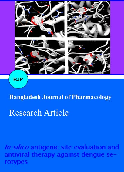
The docking of ligands to the catalytic triad of NS3-NS2B protease was performed using AutoDock 4.0 software. AutoDock is reported to be a very common docking program (Sousa et al., 2006) and is reliable (Hetényi et al., 2002). Using the software, polar hydrogen atoms were added to the enzyme and its nonpolar hydrogen atoms were merged, whereas for the ligand, nonpolar hydrogen atoms were merged and Gasteiger charges were added. All rotatable bonds of ligands were set to be rotatable. All calculation for protein-fixed ligand-flexible docking was done using the Lamarckian Genetic Algorithm (LGA) method. A population size of 150 and 10 millions energy evaluations were used for 100 search runs. The grid box with a dimension of 60 × 60 × 60 points and 0.375 Å grid spacing was used around the catalytic triad to cover the entire enzyme binding site and allow accommodated ligands to move freely. After the docking searches were completed, clustering histogram analysis was performed based on an RMSD (root mean square deviation) of not more than 1.5 Å. The best conformation was chosen from the most populated cluster with the lowest docked energy. The interactions of complex enzyme-ligand conformations, including hydrogen bond and other interactions, were analyzed using Viewerlite and UCSF Chimera software (Figure 1-4).
Figure 1: Docking Interaction of Models with ligand1 and 2. The interaction energy was calculated and the hydrogen bonds were observed
Figure 2: Docking interaction of Models with ligand 5 and 6. The interaction energy was calculated and the hydrogen bonds were observed
Figure 3: Docking interaction of Models with ligand 7 and 8. The interaction energy was calculated and the hydrogen bonds were observed
Figure 4: Docking interaction of Models with ligand 9. The interaction energy was calculated and the hydrogen bonds were observed
Results
The antigenic sites analyzed from all the serotypes were found to be highly variable. In DENV-I the sites were found to be IVGLYGNGVVTTSGTYVSPIAQAK, MRLLSPVRVP, EVQVIAVE, MAVGIVSILLSSLLKND VPLAGPLIAGGMLIACYVISG, FTVVVGDVVGILAQ and HGTVLVQVKY. It is observed that the amino acids present at the site producing highest scores were found to be A, P, V, C, G and Q for DENV I. For DENV-II, the antigenic sites were found to be EVQVLALE, MRLLSPVRVPNYNLI, QLGQVMLLVLCVTQVLM, RYLPAIVREA, TSLSVSLVLVGIVTLYLGVMVQAD, and QLGQVMLLVLCVTQVLM. The amino acids producing highest scores for DENV-II were found to be V, P and L. However DENV-III showed much conserved antigenic sequences and was found to be KYLPAIVREA, DRVIDPRRCLKPVILT, and ADRVI DPRRCLKPVILTD. Proline was found to be the most conserved antigenic site for DENV-III. The DENV-IV found to be having the most similar antigenic sites as of DENV-III. The sites were found to be IVDLMCHAT, GRVIDPRRCLKPVILT, MADLSLEKAANVQ, QLGQVMLLVLCAGQLLLM, KHMILVVVITLCAIIL GG, and GRVIDPRRCLKPVILT. The high scoring regions were found to be L, P, E and I. Hence, the antigenic site P is found to be the most conserved region among all the four serotypes, suggesting as the potent inhibition point of the ligands. The geometry and stereochemistry of the models were evaluated by using the program PROCHECK (Sousa et al., 2006). The stereochemical quality of the generated homology models as well as the crystal structure was evaluated using Ramachandran plots. Results revealed that 79.7 and 2.4% of the residues of the models are located in the most favored regions and the additional allowed regions for Model1. Similarly the residues present in favored region and disallowed regions were found to be 88.6 and 0.2, 87.1 and 11.2, 89.3 and 0.4% for Model2, Model3 and Model4 respectively. The obtained results indicate that all models possess sufficient stereochemical quality. The Phylogenetic analysis showed the high intraspecies variation in NS3 protein of all the serotypes (Figure 5). The distances within group was found to be 0.05, 1.27, 0.70 and 0.20 for DENV-I, DENV-II, DENV-III and DENV-IV respectively. The mean distances within groups showed that DENV-I has high distance related from DENV-II, DENV-III and DENV-IV. The homology models of the NS3 protein of four serotypes also showed very good variation in Ramachandran plot. The docking study of the derived compound of mycophenolic acid (4 nos.) and ribavirin (5 nos.) was done with AutoDock4.2. The binding energy obtained in the range of -0.97 to +190.03. The ligand 3 of mycophenolic acid was found to be the best drug in Model1 having the minimum binding energy -9.2 kcal/mol. The ligand 4 of the ribavirin revealed as the best inhibitor for model2 and showed the minimum binding energy of -16.5 kcal/mol and was found to be the best ligand among all the ligands studied. The number 4 ligand of the mycophenolic acid found to be second best inhibitor for model3 among all the selected ligands after the ligand4 of mycophenolic acid with binding energy -15.9 kcal/mol. The ligand1 of ribavirin was found to be the best inhibitor for model4 having minimum binding energy -10.7 kcal/mol (Figure 6).
Table V: Molecular properties of mycophenolic acid derivatives
| Sl. No | IUPAC Name | Chemical formula | Molecular weight | Log P | Structure |
|---|---|---|---|---|---|
| 1 | 6-(4-hydroxy-6-methoxy-7-methyl-3-oxo-1,3-dihydro-isobenzofuran-5-yl)-4-methyl-hex-4-enoic acicd | C17H20O6 | 320.34 | 2.55 | |
| 2 | 7-hydroxy-6(6-hydroxyamino-3-methyl-hepta-2,6-dienyl)-5-methoxy-4-methyl-3H-isobenzofuran-1one | C18H23NO5 | 333.38 | 2.63 | |
| 3 | ACMPHA | C20H26N2O5 | 374.43 | 1.91 | |
| 4 | LMPHA | C31H46N2O4 | 510.71 | ||
| 5 | 5-amino-1-(3,4-dihydroxy-5-hydroxymethyl-tetrahydro-furan-2-yl)-1H-imidazole-4-carboxylic acid amide | C9H14N4O5 | 258.23 | -3.28 | |
| 6 | 4-hydroxy-5-(3,4,5-trihydroxy-tetrahydro-furan-2-yl)-4,5-dihydro-1H-pyrazole-3-carboxylic acid amide | C8H13N3O6 | 247.21 | -3.73 | |
| 7 | 1-(3,4-dihyroxy-5-hydroxymethyl-tetrahydro-furan-2-yl)-1H-[1,2,4]triazole-3-carbozamidine | C8H13N5O4 | 243.22 | -1.61 | |
| 8 | Ribavirin 5 triphosphate | C8H12N4O14P33- | 481.12 | ||
| 9 | 1-(3,,4,5-trihydroxy-tetrahydro-furan-2-yl)-1H-[1,2,4]triazole-3carboxylic acid amide | C7H10N4O5 | 230.18 | -1.38 |
Figure 5: Binding enegry analysis from the interaction study of the ligands it was observed that the ligands 3, 8, 4 and 5 are the best inhibitors for DEN-I, DEN-II, DEN-III and DEN-IV respectively. These ligands showed the binding energy of -9.18, -16.52, -15.87 and -10.66 Kcal/mol respectively for the corresponding serotypes
Figure 6: Phylogenetic analysis of DENV I
Discussion
Dengue fever epidemics has increased numerously over the last few decades (Ligon et al., 2005) therefore Developing antiviral drug and vaccine is becoming very important due to the global threat of viral disease pandemics (Noble et al., 2010; Wang et al., 2009). The functional similarity between the NS2B/NS3 proteases from the four genetically and antigenically distinct serotypes was identified by the differences in their substrate specificity using tetrapeptide and octapeptide libraries in a positional scanning format, each containing 130,321 substrates (Guzman et al., 2010). Development of new genomic and proteomic studies coupled with computational sciences could provide the discovery of various target proteins and potential inhibitor to be developed as drugs (Li et al., 2005; Tambunan et al., 2011). The NS3 enzyme of dengue is responsible for replication of the virus. The replication complex include the NS3 nucleotide the NS3 protease and with its NS2B cofactor, the NS3 nucleotide triphosphatase. This protein serves as the potential inhibitory targets for antiviral agents since they are required for virus replication. The multifunctional C-terminal domain of NS3 encodes NTPase, helicase and RTPase activities. NTP hydrolysis is thought to provide the chemical energy required for helicase activity. There is currently no antiviral therapy available against dengue virus. In this study we generated some antiviral ligands of mycophenolic acid and ribavirin. We predicted the binding activity of the ligands against NS3 protein of Dengue virus. From the interaction study of the ligands it was observed that the ligands 3, 8, 4 and 5 are the best inhibitors for DEN-1, DEN-2, DEN-3 and DEN-4 respectively. These ligands showed the binding energy of -9.2, -16.5, -15.9 and -10.7 Kcal/mol respectively for the corresponding serotypes. To study the interaction of ligands at the inhibition point, it is necessary to know the antigenic sites of the disease causing protein. It is observed that, there is great variation in the antigenic site of the proteins. However the most conserved antigenic site found to be the aminoacid, Proline almost in every serotype. The ligands were docked into each antigenic site of the protein and the binding energy was reported. Phylogenetic analysis also suggested the typical variations in all the NS3 sequences of NCBI. Finding antigenic site of a mutated protein is the primary aim of any computer aided drug design. The current research discovered several variations in the original sequences as well as antigenic site in the NS3 protein of the 4 serotypes. The antigenic sites were targeted to block the replication activity of the virus. The antigenic sites were targeted by the derivative compounds of mycophenolic acid and ribavirin to block the replication process of the virus. Hence the current study may be useful in designing the drugs that may be synthesized in wet lab and can be used as antiviral drugs against dengue serotypes. The docking of two groups of inhibitors from mycophenolic acid and ribavirin again NS3 were carried out. In this work, the complexation energy of the docking was used as the descriptors for selecting new candidates for competitive dengue inhibitors. The antigenic sites were highly variable. The phylogenetics studies were also evaluated for finding the interspecies variation in NS3. Homologies of protein were constructed for all the serotypes. AutoDock4.2 helped in carrying out the interaction of the drugs with the protein models. Complexation energies for all the new ligand-enzyme complexes were evaluated. Detailed structural information is becoming increasingly available for the dengue NS3 proteins. Since these proteins are requested for virus infectivity and replication. Structure based computational approaches offer an attractive strategy for the discovery and optimization of dengue antiviral drugs. Moreover, these computational approaches promise and improve the effectiveness of current structure based calculations.
Conclusion
Based on the complexation energies calculated, the 3rd compound of the mycophenolic was found to be the best inhibitor for DENV I. Similarly drug4 of ribavirin, drug4 of mycophenolic acid and drug1 of ribavirin were found to be best drugs against DENV II, DENV III and DENV IV respectively the lowest and closest energies to the reference compounds.
References
Allison AC, Eugui EM. Immunosuppressive and other effects of mycophenolic acid and an ester prodrug, mycophenolate mofetil. Immunol Rev. 1993; 136: 5-28.
Bera AK, Kuhn RJ, Smith JL. Functional characterization of cis and trans activity of the flavivirus NS2B-NS3 protease. J Biol Chem. 2007; 282: 12883-92.
Bhattacharya A, Wunderlich Z, Monleon D, Tejero R, Montelione GT. Assessing model accuracy using the homology modeling automatically software. Proteins 2008; 70: 105-18.
Brinkworth RI, Fairlie DP, Leung D, Young PR. Homology model of the dengue 2 virus NS3 protease: Putative interactions with both substrate and NS2B cofactor. J Gen Vir. 1999; 80: 1167-77.
Conner CS. Ribavirin. Drug Intell Clin Pharm. 1984; 18: 137-38.
Diamond MS, Zachariah M, Harris E. Mycophenolic acid inhibits dengue virus infection by preventing replication of viral RNA. Virology 2012; 304: 211-21.
Gentry MK, Henchal EA, McCOWN LM, Brandt WE, Dalrymple JM. Identification of distinct antigenic determinants on dengue-2 virus by using monoclonal antibodies. Am J Trop Med Hyg. 1982; 31: 548-55.
Gratz NG. Emerging and resurging vector-borne diseases. Annu Rev Entomol. 1999; 44: 51-75.
Gubler DJ. Dengue and dengue hemorrhagic fever. Clin Microbiol Rev. 1998; 11: 480-96.
Guzman MG, Halstead sb, Artsob H, Buchy P, Farrar J, Gubler DJ, Hunsperger E, Kroeger A, Margolis HS, MartÃnez E, Nathan MB, Pelegrino JS, Simmons C, Yoksan S, Peeling RW. Dengue: A continuing global threat. Nature Rev Micro. 2010; S7-S16.
Halstead SB. Dengue. Lancet 2007; 370: 1644-52.
Henchal EA, Gentry MK, McCowN JM, Brandt WE. Dengue virus-specific and flavivirus group determinants identified with monoclonal antibodies by indirect immunofluo-rescence. Am J Trop Med Hyg. 1982; 31: 830-36.
Henikoff S, Henikoff JG. Amino acid substitution matrices from protein blocks. Proc Natl Acad Sci. 1992; 15: 10915-19.
Hetényi C, van der Spoel D. Efficient docking of peptides to proteins without prior knowledge of the binding site. Protein Sci. 2002; 11: 1729-37.
Koff WC, Elm JL Jr, Halstead SB. Antiviral effects of ribavirin and 6-mercapto-9-tetrahydro-2-furylpurine against dengue viruses in vitro. Antivir Res. 1982; 2: 69-79.
Li J, Lim SP, Beer D, Patel V, Wen D, Tumanut C, Tully DC, Williams JA, Jiricek J, Priestle JP, Harris JL, Vasudevan SG. Functional profiling of recombinant NS3 proteases from all four serotypes of dengue virus using tetrapeptide and octapeptide substrate libraries. J Bio Chem. 2005; 280: 28766-74.
Ligon BL. Dengue fever and dengue hemorrhagic fever: A review of the history, transmission, treatment, and prevention. Semin Pediatr Infect Dis. 2005; 16: 60-65.
Luo D, Xu T, Hunke C, Grüber G, Vasudevan SG, Lescar J. Crystal Structure of the NS3. J Virol. 2008; 82: 173-83.
Malachowska-Ugarte M, Cholewinski G, Dzierzbicka K, Trzonkowski P. Synthesis and biological activity of novel mycophenolic acid conjugates containing nitroacridine/acridone derivatives. Euro J Med Chem. 2012; 54: 197-201.
Mason PW, Zfigel MU, Semproni AR, Fournier MJ, Mason L T. The antigenic structure of dengue type 1 virus envelope and NS1 proteins expressed in Escherichia coil. J Gen Vir. 1990; 71: 2107-14.
Monath TP. Pathobiology of the flaviviruses. In: The Togaviridae and Flaviviridae. Schlesinger S, Schlesinger MJ (eds). New York, Plenum Press, 1986, 275-440.
Muhamad M, Kee LY, Rahman NA, Yusof R. Antiviral actions of flavanoid-derived compounds on dengue virus type-2. Int J Biol Sci. 2010; 6: 294-302.
Noble CG, Chen YL, Dong H, Gu F, Lim SP, Schul W, Wang QY, Shi PY. Strategies for development of dengue virus inhibitors. Antiviral Res. 2010; 85: 450-62.
Qi RF, Zhang L, Chi CW. Biological characteristics of dengue virus and potential targets for drug design. Acta Biochim Biophys Sin. 2008; 40: 91-101.
Russell PK, Nisalak A. Dengue virus identification by the plaque reduction neutralization test. J Immun. 1967; 99: 291- 96.
Sousa SF, Fernandes PA, Ramos MJ. Protein-ligand docking Current status and future challenges. Proteins 2006; 65: 15-26.
Tambunan USF, Apriyanti N, Parikesit AA, Chua W, Wuryani K. Computational design of disulfide cyclic peptide as potential inhibitor of complex NS2B-NS3 dengue virus protease. Afr J Biotechnol. 2011; 10: 12281-90.
Wang QY, Patel SJ, Vangrevelinghe E, Xu HY, Rao R, Jaber D, Schul W, Gu F, Heudi O, Ma NL, Poh MK, Phong WY, Keller TH, Jacoby E, Vasudevan SG. A small-molecule dengue virus entry inhibitor. Antimicrob Agents Chemother. 2009; 53: 1823-31.
Wheeler DL, Barrett T, Benson DA, Bryant SH, Canese K, Chetvernin V, Church DM, Dicuccio M, Edgar R, Federhen S, Feolo M, Geer LY, Helmberg W, Kapustin Y, Khovayko O, Landsman D, Lipman DJ, Madden TL, Maglott DR, Miller V, Ostell J, Pruitt KD, Schuler GD, Shumway M, Sequeira E, Sherry ST, Sirotkin K, Souvorov A, Starchenko G, Tatusov RL, Tatusova TA, Wagner L, Yaschenko E. Database resources of the National Center for Biotechnology Information. Nucleic Acids Res. 2008; 36: D13-21.