Free radical scavenging, in vivo antioxidant and hepatoprotective activity of folk medicine Trichodesma sedgwickianum
Abstract
Trichodesma sedgwickianum has been used in folk medicine possessing anti-inflammatory and hepatoprotective activity. This led us to investigate for its antioxidant and hepatoprotective potential. Different polarities extracts were subjected to polyphenolic estimation and in vitro antioxidant activity. The potential extract was tested for in vivo antioxidant and hepatoprotective activity, assessed by carbon tetrachloride-induced oxidative stress in rats. Phytochemical identification of major constituents has been carried out by HPLC, GC-MS, 1HNMR and 13C NMR. Amongst the extracts, successive ethanol extract showed higher concentration of polyphenols (25.4 ± 0.1% w/w) and in vitro antioxidant property. The in vivo antioxidant efficiency was confirmed by comparing the enzymatic level, superoxide dismutase, catalase, reduced glutathione and MDA in test group with the standard and control. Hepatoprotective effect was observed by changes in the serum enzyme level which were further supported by histological examination. Phytochemically ethanol extract contains gallic acid and catechin along with other constituents. Thus present study provides a scientific rationale for their traditional use.
Introduction
Plant kingdom with an estimated population of about 500,000 species of which only half is described so far is among the most attractive pools of bioactive natural products. Because of a range of biological activities, including antioxidant, antibacterial and antiviral effects, herbs are used as food and cosmetics preservatives and are valuable ingredients in formulation of new functional foods (Chan et al., 2007).
Antioxidants are substances when present in foods or body at low concentrations compared with that of an oxidizable substrate markedly delay or prevent the oxidation of that substrate. Antioxidants may help the body to protect itself against various types of oxidative damage caused by reactive oxygen species, which are linked to a variety of disorders including cancer, diabetes, hepatotoxicity, arthritis, and acceleration of the ageing process (Shahidi, 1997). Amongst the antioxidants, several groups of polyphenols (anthocyanins, tannins, flavanones, isoflavones, resveratrol and ellagic acid) are currently used in the industry as nutraceuticals or functional foods (Epsin et al., 2007).
Trichodesma sedgwickianum are important members of Boraginaceae family. It is cultivated mainly for its pyrrazolidine alkaloids; this species possess known anti-inflammatory, analgesic, cough reflux depressant and antidiabetic properties. Chemically it contains monocrotolin, suspinine alkaloids, hexcosane, amylin and lupeol. Traditionally paste of roots is use in treatment of swelling of joints and as an emollient. Leaves of plant are useful as a liver tonic and depurative (Kirtikar and Basu, 1935). Literature review revealed that since of potential medicinal use of plant in traditional medicine still lack of systematic biological investigations. Hence in present study plant was subjected to evaluation for their antioxidant potential and role in liver toxicity protection as well as management. T. sedgwickianum
Materials and Methods
Plant materials collection and extraction
Aerial parts of T. sedgwickianum collected in the month between August-September 2009 from Amravati District, Maharashtra (India) and it is authenticated by Prof. Bhowagaokar, VIHS, Amravati, Maharashtra, India. A voucher specimen (AMT-36) has been preserved for future reference. The dried powder material (5 kg) was extracted successively with petroleum ether, chloroform, ethanol by hot continuous method, soxhlation. While aqueous extract has been prepare by maceration technique. The solvent was evaporated to dryness under pressure using rotary flash evaporator to obtain solid extracts.
Animals
Wistar albino male rats (150–250 g), obtained from the Institute’s animal house were used. They were housed under standard laboratory conditions and were fed commercial rat feed (Lipton India Ltd., Mumbai, India) and boiled water, ad libitum. All animal experiments were carried out according to institutional animal ethical committee (Approval letter no. GCPA/IAEC/ 2011/1245).
Total phenolic content
The TP content was determined by the Folin–Ciocalteau colorimetric method (Kalaskar and Surana., 2011). The TP content was calculated from the calibration curve of gallic acid and results expressed as gallic acid equivalents.
DPPH and ABTS radical scavenging assay
ABTS and DPPH˙+ quenching ability was measured according to Kalaskar and Surana (2011). The anti-radical activity was expressed in IC50 (µg/mL).
Nitro blue tetrazolium (NBT) reduction assay
The effect of scavenging superoxide radical was determined by the nitroblue tetrazolium reduction (Fu et al., 2010). The absorbance of test sample was measured at 560 nm against blank samples.
Lipid peroxidation inhibition activity (ferric thiocyanate method)
The FTC method was adapted from of Fu et al. (2010). All measurements were made in triplicate and averaged. The inhibition rate was calculated using the equation:
[(Ac _ As)/Ac] *100 (Ac = absorbance of control; As = absorbance of sample).
In vivo antioxidant and hepatoprotective activity by CCl4-induced oxidative stress
Carbon tetrachloride-induced hepatotoxicity in rats carried out according Kalaskar and Surana (2011). The studies were carried out in double dose, 200 mg and 400 mg/kg body wt. The livers from all the animals were collected at the end of experiment, washed and used for biochemical characterization of enzymes.
Isolation and characterization
The ethanol extract was selected for isolation based on their high polyphenolic content and potential antioxidant action. Separation of phytoconstituents was carried out by column chromatography over silica gel (60-120 mesh) using varying proportion of chloroform: Methanol (90:10, 75:25, 50:50, 0:100 v/v) as eluent (Hubert et al., 2011). All fractions were combined together in to four fractions I, II, III & IV as per their TLC profile. Fraction III was re-column by gradient elution using dichloromethane: Methanol (90:10, 75:25: 50:50, 25:75 v/v) (Kumaraswamy et al., 2010), as it shows presence of only three compounds in TLC. All fractions obtained in re-column were monitored using silica gel TLC. These fractions were combined and concentrated in rotary vacuum evaporator. The major single crystalline compound was obtained and named as TRS-I. Isolated compound was characterized by UV, HPLC, GC-MS and 1H and C13 NMR. The UV spectrum was recorded on 1700 UV-Visible spectrometer.
1HNMR and 13C NMR spectra were recorded on a Varian-500 and Varian-100 MHZ respectively at IIT Powai, Mumbai India. The 1HNMR and 13C NMR spectra were recorded using DMSO as solvent and TMS as an internal standard. The GC-MS was recorded at high resolution on a mass spectrometer (Perkin Elmer Autosystem XL with Turbomass) at Saif, IIT Powai, Mumbai India and data are given in m/z value.
The above compounds were also subjected to the HPLC analysis by using the standard marker (Sigma-Aldrich Chemie, Steinheim, Germany). Mobile phase consisted of acetonitrile: Water (80:20) mixture with flow rate 0.3 mL/min, at 25ºC was employed for TRS-I. Detection wavelength was set at 280 nm. Data was acquired and analyzed using chromquest version 3.0 software.
Statistical analysis
Results were expressed as mean ± SEM. Data were analyzed using one-way analysis of variance (ANOVA) followed by Dunnett’s test. Value of p<0.05 was considered to be statistically significant.
Results
Phenolic compounds are considered to be the major contributors to the antioxidant capacity of plants. Some of diverse biological activities of plant may also be related to their antioxidant activity (Chung et al., 1998). The ethanol extract of T. sedgwickianum was found to be possessing highest concentration (% w/w) of phenolics 25.4 ± 0.1 amongst the other extracts, 12.1 ± 0.1, 14.2 ± 0.2, and 19.3 ± 0.3 in petroleum ether, chloroform and acetone extract, respectively.
Antioxidant activity of different extracts was evaluated by free radical scavenging by ABTS and DPPH, superoxide scavenging and lipid peroxide inhibition. The concentration of each extract required to inhibit each radical by 50% (IC50) is shown in Table I. The result revealed that the ethanol extract exhibits the most robust radical-scavenging activity among the extracts after standard ascorbic acid.
Table I:IC50 value for various extracts of in vitro antioxidant assay
| Sample | IC50 value (µg/mL) | |||
|---|---|---|---|---|
| DPPH | ABTS | Lipid peroxide | Superoxide | |
| Petroleum ether | 56.9 ± 1.0 | 47.1 ± 0.3 | 78.2 ± 0.3 | 78.3 ± 0.2 |
| Chloroform extract | 34.0 ± 0.1 | 34.4 ± 0.9 | 48.4 ± 0.2 | 41.3 ± 0.5 |
| Ethanol extract | 32.5 ± 1.1 | 24.1 ± 0.3 | 38.5 ± 0.1 | 29.5 ± 0.1 |
| Acetone extract | 41.3 ± 0.2 | 39.8 ± 0.4 | 55.3 ± 0.4 | 64.4 ± 0.9 |
| Ascorbic acid | 20.6 ± 0.3 | 19.8 ± 0.2 | 23.5 ± 0.4 | 20.2 ± 0.2 |
| Values are the mean ± SEM; n = 3 | ||||
Based on our in vitro antioxidant assays, ethanol extract was chosen as the most potent extract and subsequently it was used to evaluate in vivo antioxidant and hepatoprotective potential.
For the evaluation of in vivo antioxidant and hepatoprotective activity, carbon tetrachloride-induced hepatotoxicity model was extensively used. The in vivo antioxidant properties of ethanol extract in liver are presented in Table II. Compared with the control group, the carbon tetrachloride-intoxicated animals exhibited a significant decrease in superoxide dismutase, catalase, and reduced glutathione levels, together with a significant increase in the level of MDA in liver (p<0.05). These changes were significantly reversed in a dose dependent manner upon treatment with SEE, or the standard treatment silymarin (Halliwell and Gutteridge, 1990)
Table II:Effects of ethanol extract of Trichodesma sedgwickianum on liver superoxide dismutase, catalase, MDA, and reduced glutathione in carbon tetrachloride-intoxicated rats
| Group | Liver | |||
|---|---|---|---|---|
| Superoxide dismutase (U/mg protein) |
Catalase (U/mg protein) |
MDA (nmol/mg protein) |
Reduced glutathione (mg/g protein) |
|
| Normal | 335.9 ± 24.6 | 62.1 ± 12.7 | 2.5 ± 0.5 | 5.9 ± 1.1 |
| Carbon tetrachloride | 154.7 ± 40.6a | 33.1 ± 6.1a | 5.8 ± 1.3a | 2.4 ± 0.7a |
| Silymarin | 274.0 ± 45.9b | 51.8 ± 13.3c | 3.8 ± 0.4c | 4.7 ± 1.6c |
| Carbon tetrachloride + Ethanol extract (200 mg/kg) | 223.9 ± 45.8d | 40.3± 9.6c | 2.9 ± 0.5c | 4.8 ± 0.6d |
| Carbon tetrachloride + Ethanol extract (400 mg/kg) | 270.0 ± 36.7c | 54.0 ± 13.3b | 3.1 ± 0.7c | 4.4 ± 1.1d |
| Values are the mean ± SEM, n = 6; ap˂0.01 when compared with control; bp˂0.01 when compared with toxicant; cp˂0.05 when compared with toxicant; dp˃0.05 when compared with toxicant | ||||
Carbon tetrachloride-induced a marked increase in the serum level of SGOT, SGPT, ALP, Total protein, total bilirubin and direct bilirubin as compared to normal controls indicating liver damage. Pre-treatment of the rats with ethanol extract (200 and 400 mg/kg) prior to carbon tetrachloride administration caused a significant change in the values of SGOT, SGPT, alkaline phosphatase, total protein and bilirubin) in a dose dependent manner. However, ethanol extract (400 mg/kg) showed highest hepatoprotective activity (Table III) almost comparable to the silymarin (100 mg/kg) treated group.
Table III:Effects ethanol extract on rat serum parameters after carbon tetrachloride administration
| Group | SGOT (IU/L) |
SGPT (IU/L) |
Alkaline phosphatase (IU/L) |
TB (mg/dL) |
Direct bilirubin (mg/dL) |
Total protein (g%) |
|---|---|---|---|---|---|---|
| Normal | 55.2 ± 1.6 | 49.2 ± 1.3 | 92.6 ± 2.5 | 1.0 ± 0.0 | 0.4 ± 0.1 | 7.3 ± 0.2 |
| Carbon tetrachloride | 177.1 ± 3.8a | 166.3 ± 4.2a | 212.6 ± 6.1a | 5.7 ± 0.1a | 1.1 ± 0.0a | 5.0 ± 0.2a |
| Silymarin | 68.4 ± 1.8b | 55.0 ± 2.4b | 91.8 ± 2.9b | 1.9 ± 0.1b | 0.5 ± 0.0b | 6.9 ± 0.1b |
| Carbon tetrachloride + Ethanol extract (200 mg/kg) | 128.2 ± 2.5b | 96.6 ± 2.0b | 139.0 ± 1.7b | 2.6 ± 0.1b | 0.7 ± 0.1b | 6.7 ± 0.1c |
| Carbon tetrachloride + Ethanol extract (400 mg/kg) | 91.3 ± 2.1b | 69.07 ± 1.6b | 98.6 ± 1.4b | 1.4 ± 0.0b | 0.6 ± 0.0b | 6.7 ± 0.1b |
| Values are the mean ± SEM, n = 6; ap˂0.01 when compared with control; bp˂0.01 when compared with toxicant; cp˂0.05 when compared with toxicant | ||||||
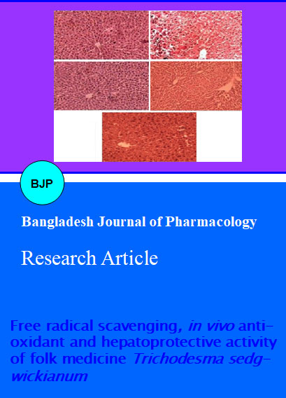
The hepatoprotective effect of ethanol extract was also supported by histological examination of the liver tissue of control and treated animals. The histological architecture of carbon tetrachloride-treated liver section showed marked massive fatty changes, necrosis, ballooning degeneration and the loss of cellular boundaries. However necrosis was not observed in any groups which indicate that sufficient hepatotoxicity does not seems to have developed in the animals so as to cause the necrosis of liver (Figure 1).
Figure 1:Histological architecture of liver of control and experimental rats
A: Normal control; B: Carbon tetrachloride; C: Silymarin treated; D: Ethanol extract of Trichodesma sedgwickianum-treated (200 mg/kg), E- Extract-treated (400 mg/kg)
Phytochemical characterization
TRS-I: 1HNMR (Chemical shift in δ ppm) : 5.925 (1H, d, J=2.1 Hz, H-8), 5.858 (1H, d, J=2.1 Hz, H-6), 4.55 (1H, d, J=7.8 Hz, H-2), 3.968 (1H, m, H-3), 2.821 (1H, dd, J=16.0, 1.6 Hz, H-4b), 2.495 (1H, dd, J=16.0, 8.6 Hz, H-4a), 6.834 (1H, d, J=1.8 Hz, H-2′), 6.753 (1H, d, J=8.1 Hz, H-5′), 6.704 (1H, dd, J=8.1, 1.8 Hz, H-6′).
13C NMR (DMSO, Chemical shift in δ ppm): δ28.55 (C-4), δ 68.85 (C-3), δ 82.88 (C-2), δ 96.37 (C-6), δ100.9 (C-10), δ 115.32 (C-2´), δ116.18 (C-5´), δ 120.13 (C-6´), δ 132.25 (C-1´), δ146.29 (C-4´), δ156.96 (C-9), δ157.6 (C-5), δ157.85 (C-7).
GC-MS: 290.50, 281.11, 207.07, 167.21, 153.19, 139.17, 125.14, 111.12, 97.10, 83.09, 71.09, 57.07.
The melting point of TRI-I was 175-177°C and UV λmax value was 276 nm. The 1H-NMR spectrum exhibited signals of a flavan-3-ol framework confirmed by proton signals at d δ4.57 (d, J=7.2 Hz), δ3.93 (m), δ2.49 (dd, J=7.8, 16.5 Hz), δ2.821 (m). The aromatic signals at d δ6.704 (d, J=8.1Hz, H-6'), δ6.709 (d, J=8.1Hz, H-5') and δ6.74 (d, J=2.2Hz, H-2'); the appearance of a one-proton aromatic at d δ5.92 (s, H-8) indicated the occurrence of a tri substitution system in the flavan A-ring. Since the proton signal as a doublet (J= 7.2 Hz) at δ 4.57, the relative configuration of the C-ring of TRI-I was the same as that of catechin (2, 3-trans) (Liu et al., 2005). 13C-NMR data indicated that TRI-I possessed nineteen sp2 carbons and five sp3 carbons. In addition to fifteen signals similar to those of catechin, 13CNMR data revealed the presence of a phenyl propanoid (C6-C3) unit with an aromatic 3, 4-dihydroxy system, which was linked with the carbonyl β-position through a carbon-carbon bond.
Mass spectrum showed a parent molecular ion [M+] peak at m/z 291 which corresponds to the molecular formula C15H14O6. The mass spectrum consisted of daughter ions at m/z 139, 125, and 167. The fragment ions peaks m/z 139 corresponds to A-ring resulted from retro-Diels-Alder ï¬ssion (RDA) of the heterocyclic ring system, and the fragment ion m/z 125 resulted from cleavage between C2–C3 and O–C2 of the pyran ring. Another main fragment ion at m/z 167 might result from cleavage between C4–C5 and O–C2 of the pyran ring (Hye et al., 2009).
These above physiochemical and spectroscopic assignments are in good agreement for the structure of catechin which was further confirmed by literature (Kuhnle et al., 2000).
The HPLC analysis of the ethanol extract has shown the presence of catechin in extract when compared to the markers (Figure 2). The retention time of catechin has been found to be 2.6.
Figure 2: HPLC analysis of ethanol extract of Trichodesma sedgwickianum CA1- Chromatogram of standard catechin; CA2- Chromatogram of catechin in sample
Discussion
In the presence of T. sedgwickianum extracts cause inhibition of DPPH radical indicates high antioxidant capacity of extracts observed from IC50 value. Considering the fact that quenching properties were obtained from the ethanol extract greater amongst the extract and comparable to the standard ascorbic acid. Scavenging of DPPH radical is related to the inhibition of lipid peroxidation. Similar results were obtained in ABTS assay. ABTS. + is a blue chromophore produced by the reaction between ABTS and potassium persulfate. Addition of the extracts to this pre-formed radical cation reduced it to ABTS. The results were compared with the standard ascorbic acid and the IC50 value demonstrates that ethanol extract is a potent antioxidant amongst the extracts. Inhibition of DPPH and ABTS radical clearly indicates its direct role in trapping free radicals by donating hydrogen atom or electron (Kaviarasan et al., 2007).
The superoxide anion radical (▪O‾2) is the most common free radical generated in vivo. Superoxides are produced from molecular oxygen due to oxidative enzymes of body as well via non-enzymatic reaction such as autoxidation by catecholamines. The scavenging activity towards the superoxide radicals (O2.-) is measured in terms of inhibition of generation of O2.-. In present study, superoxide radical reduces NBT to blue colored formosan that is measured at 590 nm. The result shows that ethanol extract and ascorbic acid has potent scavenging activity with increasing percentage inhibition. The probable mechanism of scavenging the superoxide anions may be due to the inhibitory effect of extracts towards generation of superoxide in the in vitro reaction mixture. Suppression of ▪O‾2 radical in presense of ethanol extract was observed consequences (Li et al., 2010).
The lipid peroxidation is accelerated when free radicals are formed as the results of losing a hydrogen atom from the double bond in the structure of unsaturated fatty acids. Scavenging of free radicals is one of the major antioxidant mechanisms to inhibit the chain reaction of lipid peroxidation (Donfack et al., 2011).
Synergistic interactions amongst the various antioxidative components in the SEE of T. sedgwickianum might be responsible for the relatively high values of antioxidant activity.
Carbon tetrachloride is accumulated in hepatic parenchyma cells and metabolized to the CClâ–ª3 (Recknagel, 1993). CCl3â–ª radical reacts very rapidly with oxygen to yield a highly reactive CCl3OOâ–ª. These radicals react with proteins and lipids. They remove hydrogen atoms from unsaturated lipids thus initiating lipid peroxidation, which causes loss of integrity of cell membranes and damage to hepatic tissue (Donfack et al., 2011). The enzymes reduced glutathione, superoxide dismutase and catalase are key antioxidant enzymes that protect against oxidative stress and tissue-damage (Halliwell and Gutteridge, 1990).
These enzymes are critical for defence mechanisms against the harmful effects of reactive oxygen species (ROS) and free radicals in biological systems. The superoxide dismutase converts superoxide radicals (O2) into H2O2 and O2, thus participating with other antioxidant enzymes, in the enzymatic defence against oxygen toxicity. Catalase is a key component of the antioxidant defence system. Inhibition of this protective mechanism results in enhanced sensitivity to free radical induced cellular damage. The decrease of catalase may result in a lot of deleterious effects due to the accumulation of superoxide radicals and hydrogen peroxide (Srinivasan et al., 2007). Reduced glutathione content was another important parameter that revealed oxidative damage in both liver and kidney. Reduced glutathione constitutes the first line of defence against free radicals. Reduction in liver reduced glutathione activity in carbon tetrachloride-treated rats as observed in this study indicates the damage to the hepatic cells. Lipid peroxidation is an autocatalytic process, which is a common consequence of cell death. This process may cause peroxidative tissue damage in inflammation, cancer and toxicity of xenobiotics and aging (Halliwell 1994). MDA is a cytotoxic product that is a hallmark of lipid peroxidation. The fact that extract treatment reduced elevated MDA and increased levels of superoxide dismutase, catalase, and reduced glutathione, indicated that it prevent the peroxidation of lipids by carbon tetrachloride.
The enzymes SGPT, SGOT, ALP, TP, TB and DB are originally present in high concentrations in the cytoplasm. When liver cells are in injury, these enzymes leak into the blood stream and manifest significantly elevated serum levels. And the extent of liver damage is in conformity with the elevated serum levels of these enzymes. Alkaline phosphate is the prototype of these enzymes that reflects the pathological alteration in biliary flow (Ploa and Hewitt, 1989). Carbon tetrachloride induced elevation of this enzymatic activity in the serum is in line with high level of serum bilirubin content. The ethanol extract induced suppression of the increased ALP activity with the concurrent depletion of raised bilirubin suggest the possibility of the extracts to have ability to stabilize biliary dysfunction in rat liver during hepatic injury with carbon tetrachloride. In this study the ethanol extract demonstrated hepatoprotective activity by reducing the carbon tetrachloride-induced elevated level of SGPT, SGOT, ALP, TB and SB. Thus, administration of ethanol extract revealed potent hepatoprotective activity against the toxic effect of carbon tetrachloride which was also supported by histological studies.
The phytochemical analysis has revealed the presense of high phenolic compounds in the ethanol extract. The identity of isolated constituent from ethanol extract has been established as a catechin by spectroscopic analysis. Perhaps catechin and other related phenolic compounds present in ethanol extract may be contributed for its observed antioxidant activity.
The antioxidant and free radical scavenging property of catechin was previously reported (Tanigawa et al., 2007). Catechin causes the inhibition of nuclear factor-E2-related factor 2 (Nrf2) which was the principle determining factor for the antioxidant property (Tanigawa et al., 2007).
Conclusion
T. sedgwickianum is a potential source for antioxidant molecules. Strong antioxidant properties of this plant provide the rational for use of plant in many disorders where free radical play significant role such as inflammation, hepatitis or cancer.
Ethical Issue
All animal experiments were carried out according to institutional animal ethical committee (Approval letter No.
GCPA/IAEC/ 2011/1245).
References
Chan EWC, Lim YY, Omar M. Antioxidant and antibacterial activity of leaves of Etingera species (Zingiberaceae) in Peninsular Malaysia. Food Chem. 2007; 104: 1586-93.
Chung KT, Wong TY, Wei CI, Huang YW, Lin Y. Tannins and human health: A review. Crit Rev Food Sci Nutr. 1998; 38: 421-64.
Donfack HJ, Kengap RT, Ngameni B, Chuisseu PDD, Tchana AN, Buonocore D, Ngadjui BT, Moundipa PF, Marzatico F. Ficus cordata Thunb (Moraceae) is a potential source of some hepatoprotective and antioxidant compounds. Pharmacologia 2011; 2: 137-45.
Espin JC, Garcia-Conesa MT, Tomás-Barberán FA. Nutraceuricals: Facts and fiction. Phytochemistry 2007; 68: 2986-3008.
Fu W, Chen J, Cai Y, Lei Y, Chen L, Pei L, Zhou D, Liang X, Ruan J. Antioxidant, free radical scavenging, anti-inflammatory and hepatoprotective potential of the extract from Parathelypteris nipponica (Franch. et Sav.) Ching. J Ethnopharmacol. 2010; 130: 521-28.
Halliwell B, Gutteridge JMC. Role of free radicals and catalytic metal irons in human disease: An overview. Method Enzymol. 1990; 186: 1-85.
Halliwell B. Free radicals, antioxidants and human diseases; curiosity, cause, or consequence? Lancet 1994; 344: 721-24.
Hye MA, Taher MA, Ali MY, Ali MU, Zaman S. Isolation of (+)-catechin from Acacia catechu (Cutch Tree) by a convenient method. J Sci Res. 2009; 1: 300-05.
Kalaskar MG, Surana SJ. Free radical scavenging and hepatoprotective potential of Ficus microcarpa L. fil. bark extracts. J Nat Med. 2011; 65: 633-40.
Kaviarasan S, Naik GH, Gangabhagirathi R, Anuradha CV, Priyadarsini KI. In vitro studies on antiradical and antioxidant activities of fenugreek (Trigonella foenum graecum) seeds. Food Chem. 2007; 103: 31–37.
Kirtikar KR, Basu BD. Indian Medicinal plants. 2nd ed. India, International Book Distributors, 1935.
Kuhnle G, Spencer JPE, Schroeter H, Shenoy B, Debnam ES, Srai SK, Rice-Evans C, Hahn U. Epicatechin and catechin are o-methylated and glucuronidated in the small intestine. Biochem Biophys Res Commun. 2000; 277: 507-12.
Kumaraswamy MV, Raghavendra MP, Satish S. Antioxidant and anti-inflammatory activity of Woodfordia fructicosa Kurz. J Pharm Res. 2010; 3: 1492-95.
Li R, Chen WC, Wang WP, Tian WY, Zhang XG. Antioxidant activity of Astragalus polysaccharides and antitumour activity of the polysaccharides and siRNA. Carbohydr Polym. 2010; 82: 220-44.
Liu CZ, Yu JC, Zhang XZ, Wang T, Han JX. On changes of activity of antioxidases in hippocampus of rats with multi-infarct dementia and the intervention effects of acupuncture. Chin J Tradit Chin Med Pharm. 2005; 20: 724-26.
Ploa GL, Hewitt WR. Detection and evaluation of chemically induced liver injury. In: Principle and Methods of Toxicology. Wallace HA (ed). 2nd ed. New York, Raven Press, 1989, pp 399-628.
Recknagel RO. A new direction in the study of carbon tetrachloride hepatotoxicity. Life Sci. 1983; 33: 401-08.
Shahidi F. Natural antioxidants: Chemistry, health effects and applications. 4th ed. USA, AOCS Press, 1997, pp 366-69.
Srinivasan R, Chandrasekar MJN, Nanjan MJ, Suresh B. Antioxidant activity of Caesalpinia digyna root. J Ethnopharmacol. 2007; 113: 284-91.
Tanigawa S, Fujii M, Hou DX. Action of Nrf2 and Keap1 in ARE mediated NQO1 expression by quercetin. Free Radic Biol Med. 2007; 42: 1690-703.
Zhou D, Ruan J, Cai Y, Xiong Z, Fu W, Wei A. Antioxidant and hepatoprotective activity of ethanol extract of Arachniodes exilis (Hance) Ching. J Ethnopharmacol 2010; 129: 232-37.